|
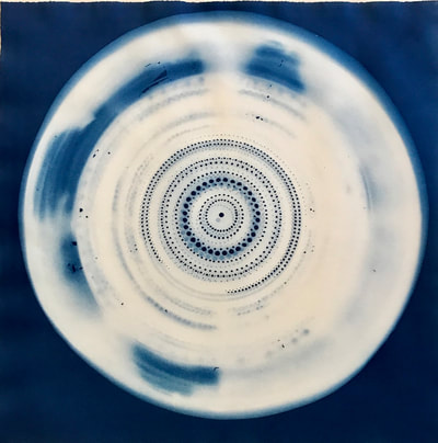
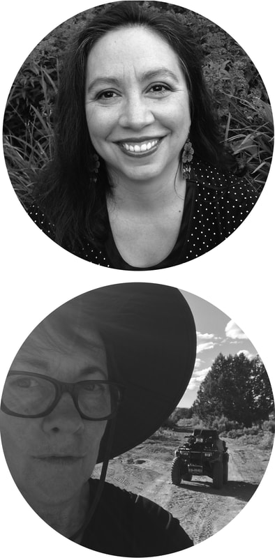
Montse When I first learned about the Bridge program, I was very intrigued and interested. The idea that a scientist and artist have a collective experience was inspiring. I am a scientist (Chemist) that loves and appreciates art, however due to various reasons I have not been able to dedicate time to nurture this interest. I have always believed that artist and scientist have a lot of common and when I began I was very excited for this opportunity. Jo Yarrington has been an amazing partner; her insight has been invaluable, I have learned a lot about how she sees the world which has enlighten me in numerous ways. One of the most valuable lesson learned was: step back and observe, no thought, just contemplate. One of my main topics of interest as a researcher is tea and coincidently Jo has used tea as an inspiration for some of her art creations. Very early on, we dive in to explore tea from two very different perspectives, thanks to Jo’s creative insight, I discovered many new things about tea that I was unaware of. For example, I learned that in order to prepare some kinds of tea, yeast is used in the process. Also I learned that some yeast and bacteria can grow in tea, which is very intriguing mostly because tea is known to have antibacterial properties. In the future I would like to explore which types of yeast and bacteria are likely to live in dry tea samples. This partnership also inspired me to change my organic chemistry classes to involve an art project. In one class the students will prepare cyanotypes, and in another class the students will design an Art in Chemistry mobile exhibit. This collaboration has helped me to find time to nurture my interest in art, specifically watercolor. The partnership between Jo and me is still ongoing; one of our future projects includes a Sci-Art exhibition at Kettering University. I have had a wonderful time being part of the Bridge program. In short, this experience has given me invaluable knowledge and has open many new exiting opportunities that I will pursue in the future. I would like to thank Julia Buntaine Hoel, Kate Schwarting from the SciArt center, as well as Dean Laura Vosejpka for all of their support and help during this residence. Jo
Although the Bridge Virtual Residency was challenging at times due to other residencies and international travel for research on concurrent projects during my sabbatical, I am extremely grateful for the opportunity to be part of the 2018 SciArt Bridge teams. In a mutually supportive and engaging collaboration with Montseurrat Rabago-Smith, our conversations and blog entries helped to expand my perspective on how art and science can exist in a dynamic framework, how they each employ analytical thinking, intense observation, and structured inquiry. Both disciplines involve processes that balance creative play and exploration with a focused purpose. I am excited to see how our work together involving materials such as tea, wax, molds, paper and alternative photographic methods such as Cyanotypes and microscope-based imagery will evolve as we continue to investigate a shared concern with environmental issues and challenges. From late February through September 2019, we have planned a series of interactions - multiple workshops and extended dialogues with other colleagues at our perspective universities and an exhibition of our collective results. This exhibition, titled The Bridge: Collaborative Work by Montseurrat Rabago-Smith and Jo Yarrington, opens this April at Kettering University Humanities Art Center and Gallery. The Bridge Virtual Residency program has been a timely gift, offering an opportunity to have a deep and meaningful dialogue across disciplines that will be affecting my teaching when I return to the university in the Fall of 2019. Although I have used a lab-like approach for projects and assignments, I plan on employing more interdisciplinary, experimental and process-based methods to aid in developing a conceptually strong hypothesis as students move toward the final completion of their artwork. Students will be asked to consider visual language across disciplines to articulate and evoke a more inclusive meaning. I would like to thank Julia Buntaine Hoel, Kate Schwarting from the SciArt center, as well as Dr. Richard Greenwald, Dean of the College of Arts and Sciences at Fairfield University, for all of their support and help during this Residency. Project plan for proposed University exchange, exhibition, lectures and interdisciplinary workshops February Develop collective exhibition
March Montse visiting Fairfield University
April Opening Reception at Kettering University Humanities Art Center and Gallery
May Jo visiting Kettering University
June Mid-exhibition reception
0 Comments
Montse Writing this blog has been a bittersweet experience. I have had a wonderful time being part of the Bridge program. It has helped me to become a better scientist as well as a better person. Jo Yarrington has been an amazing partner, her insight has been invaluable. Even though our residence is over, Jo and I will continue to collaborate. In the future, we plan to conduct a Sci-Art exhibition at Kettering University. This program has catalyzed the incorporation of art in my classes. I would like to thank Julia Buntaine Hoel, Kate Schwarting from the SciArt center, as well as Dean Laura Vosejpka for all of their support and help during this residence. Jo
Montse and I are continuing to plan for her visit to my university now scheduled for early February, my visit to her university first as preparation in later February then in May to work with her students and our exhibition. She is in Mexico and I am back in Paris so it has become a truly international virtual Residency. We have been discussing whether to start an early progressive installation in her university gallery and add materials as we develop work up to and including the workshop. I will have another work in Paris to finish up some of the art and science combined exhibitions that had been of interest earlier and that I didn’t have time to attend. I look forward to writing a summary of our collaboration in January. It certainly has been productive one so far and I am excited to see what might develop when we work together with her students. Jo
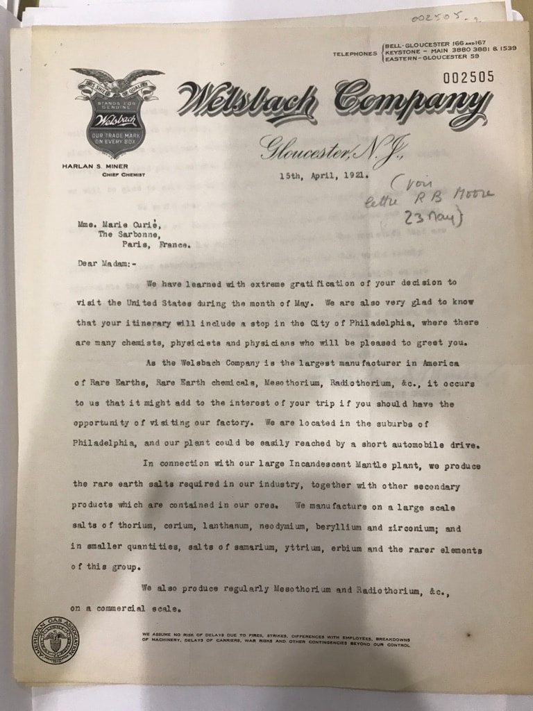
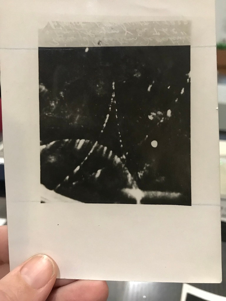
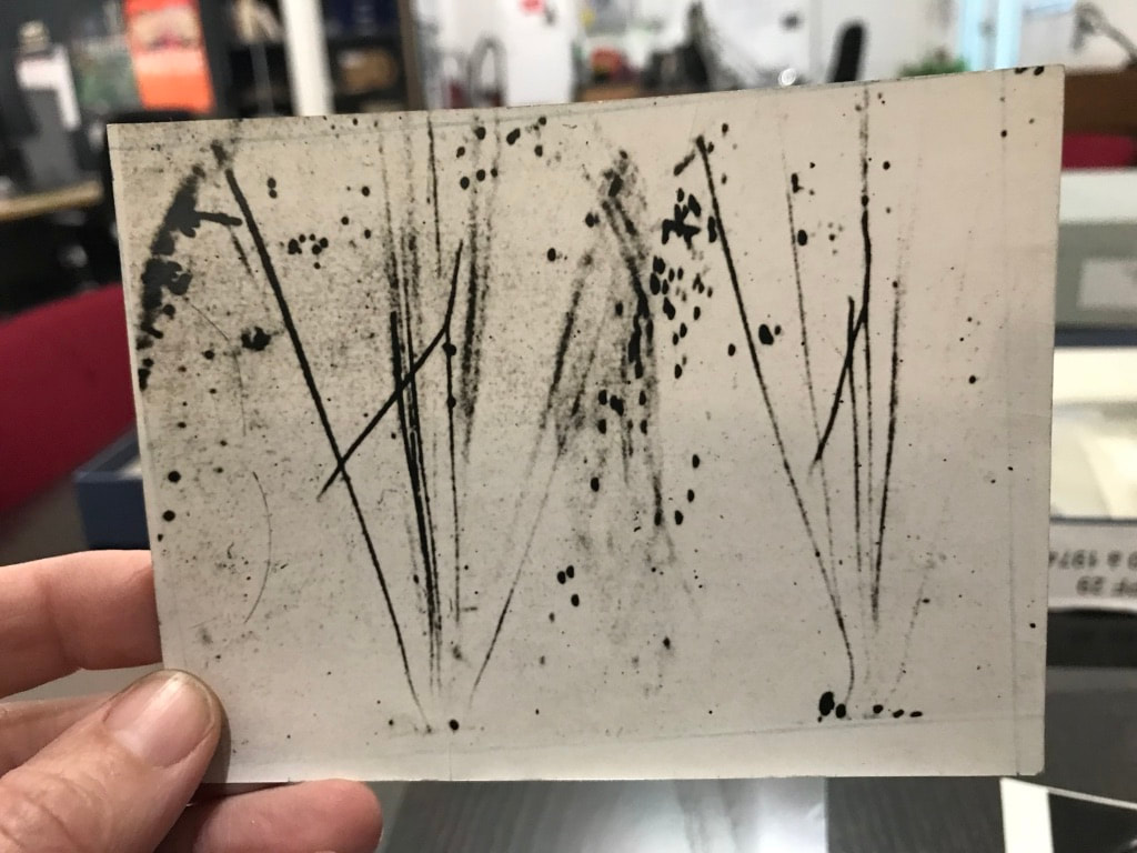
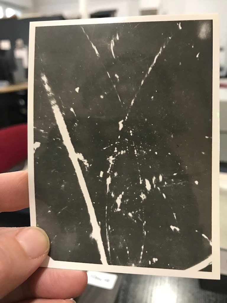
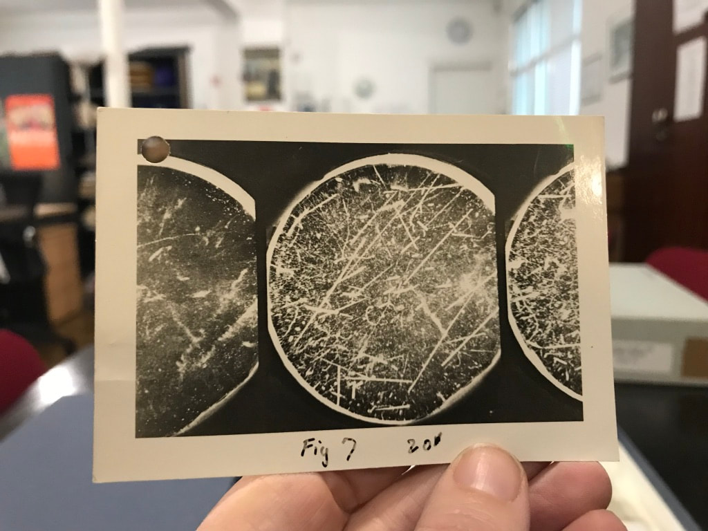
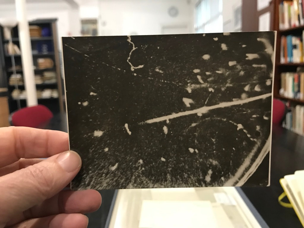
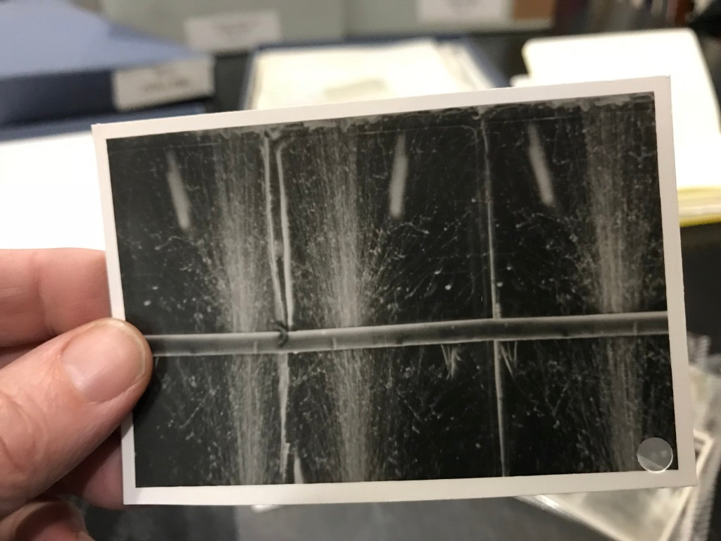
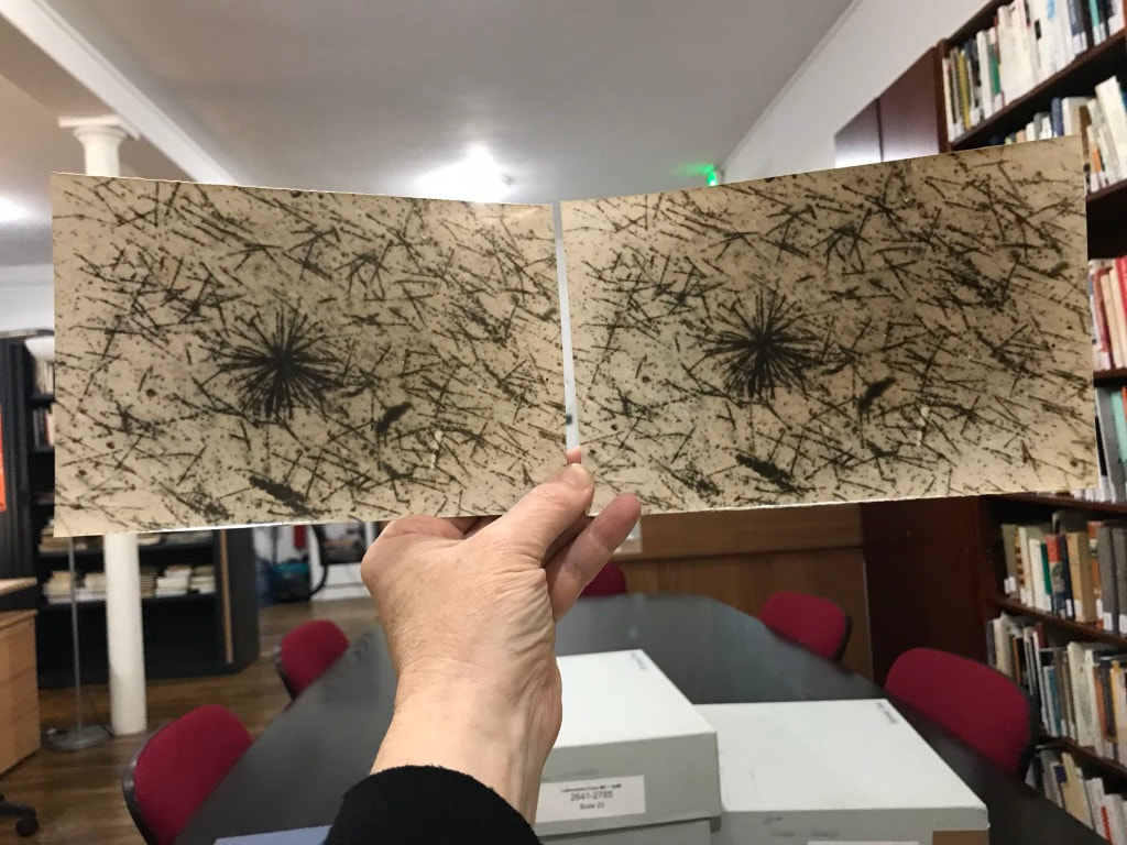
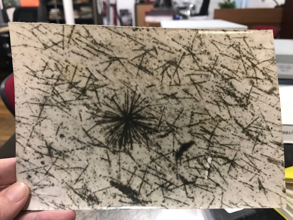
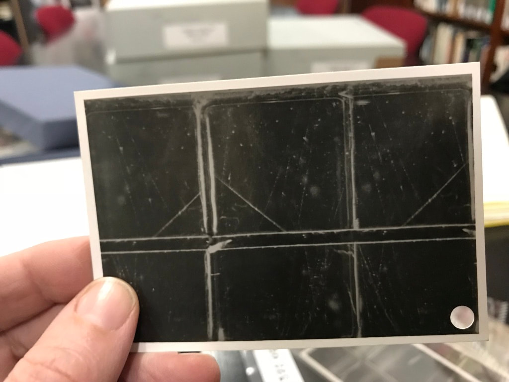
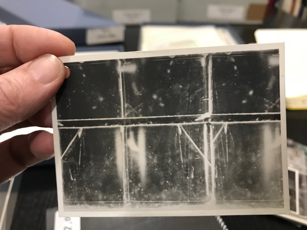
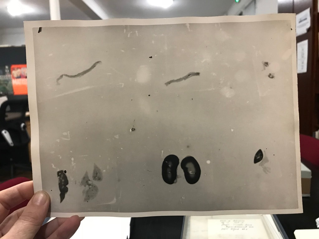
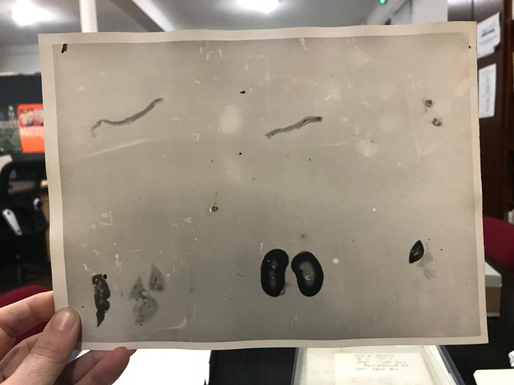
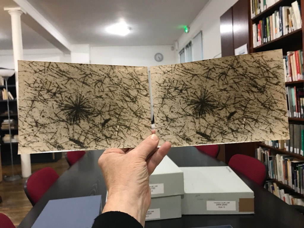
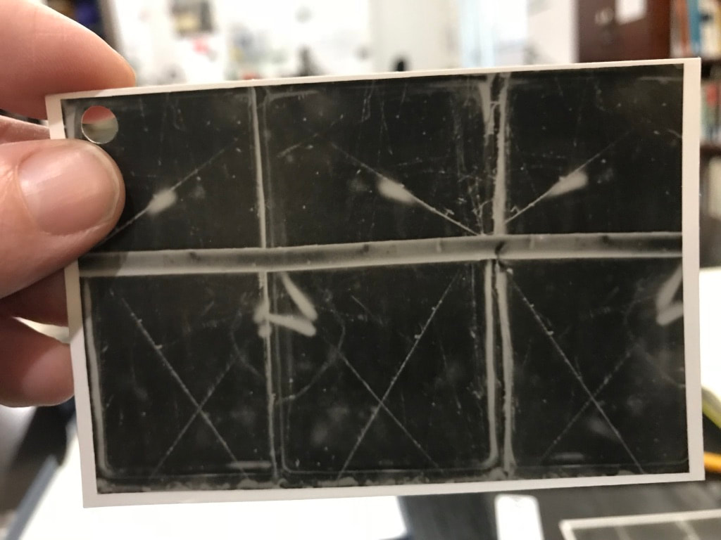
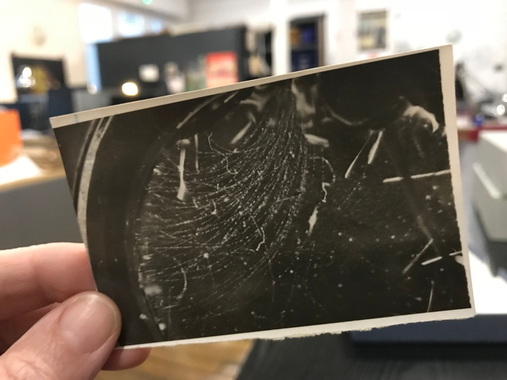
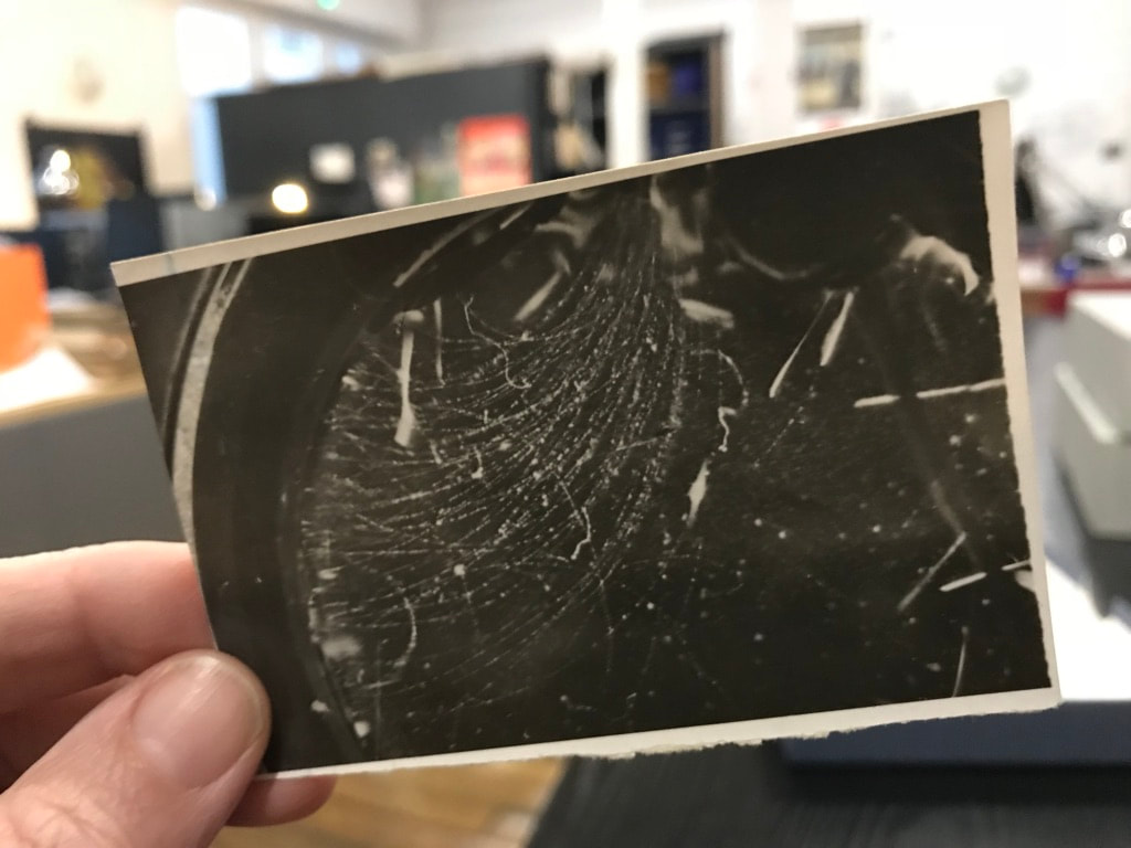
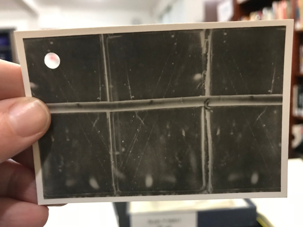
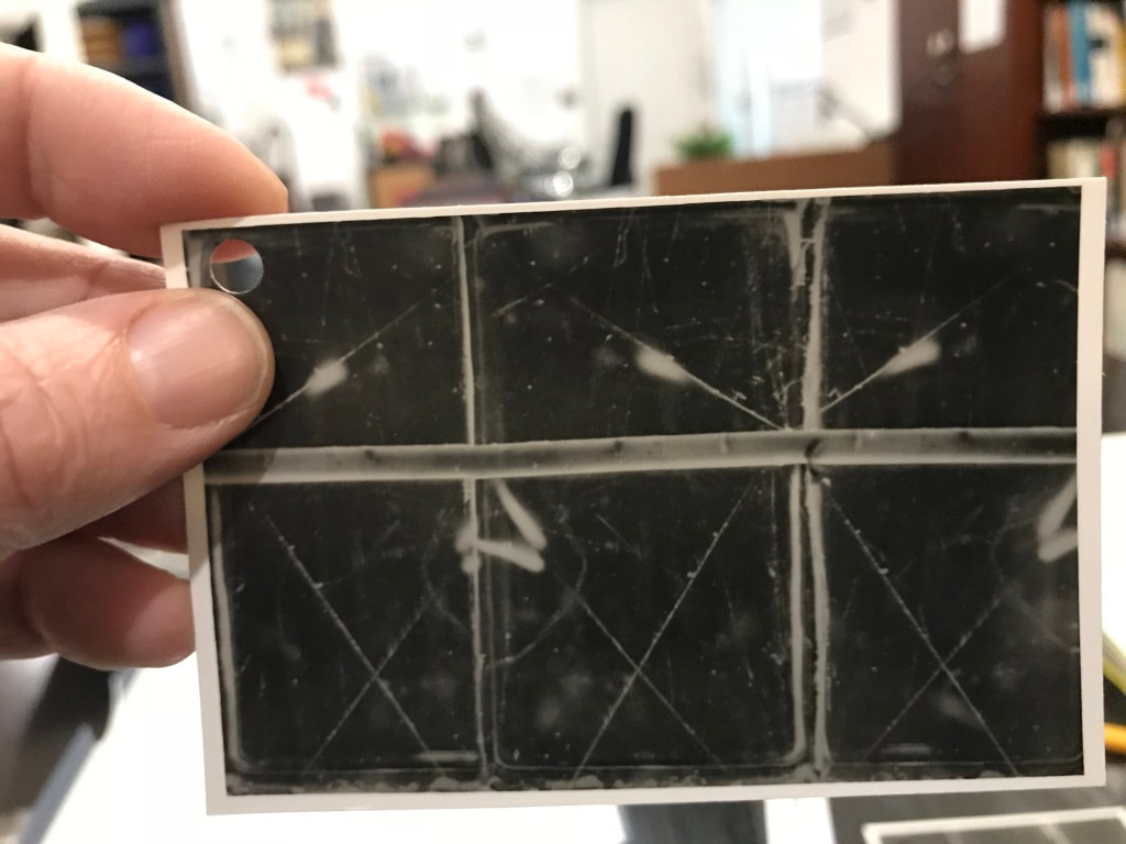
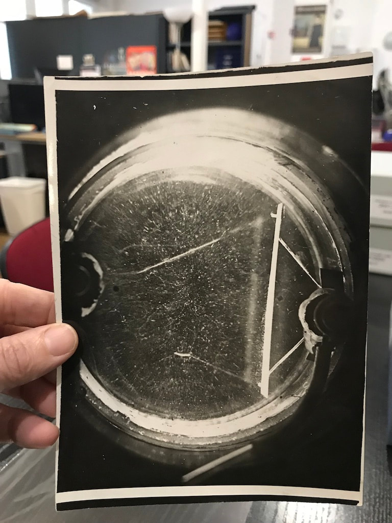
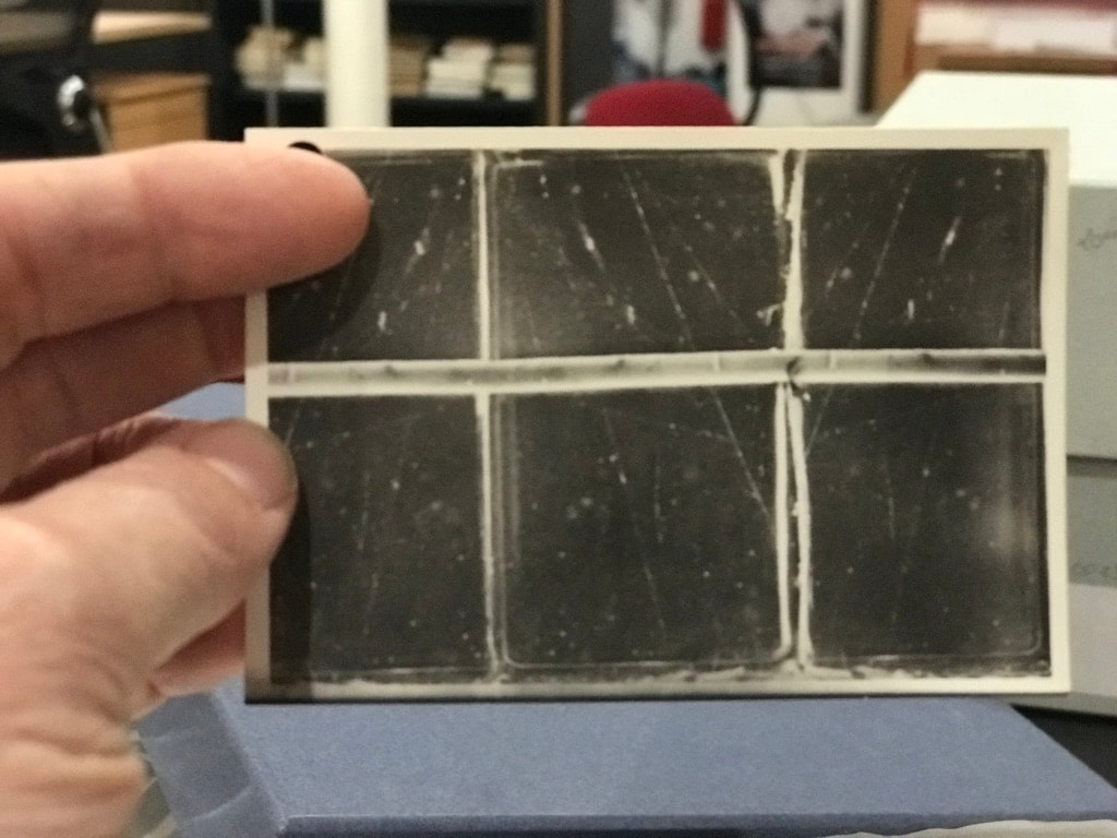
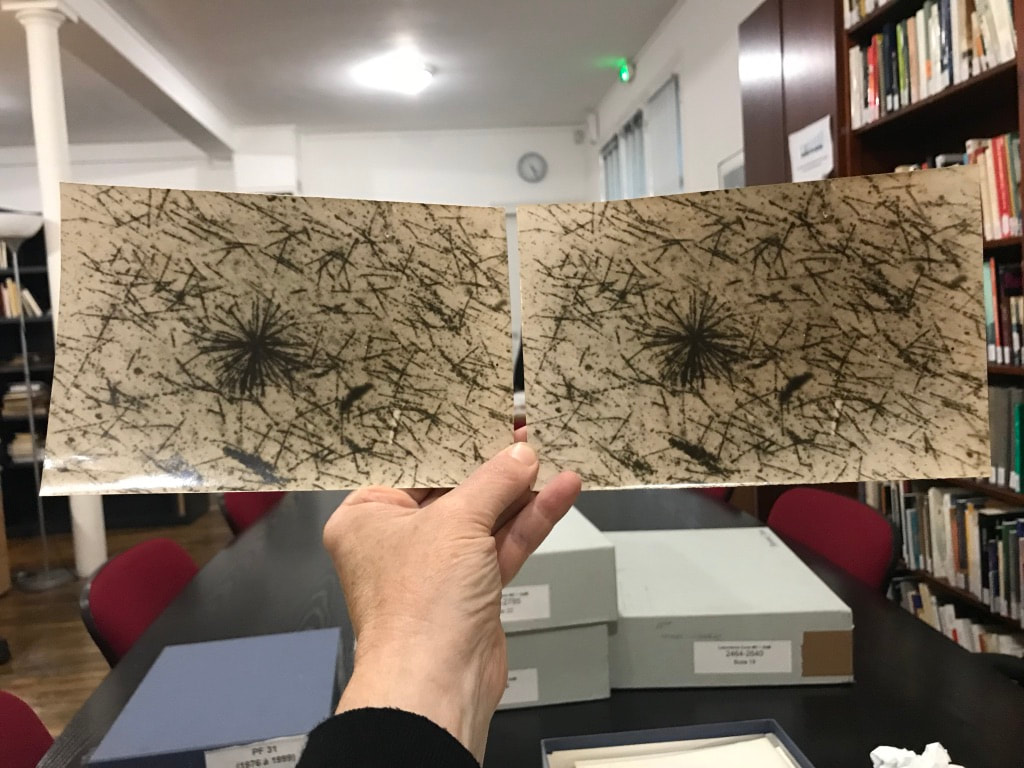
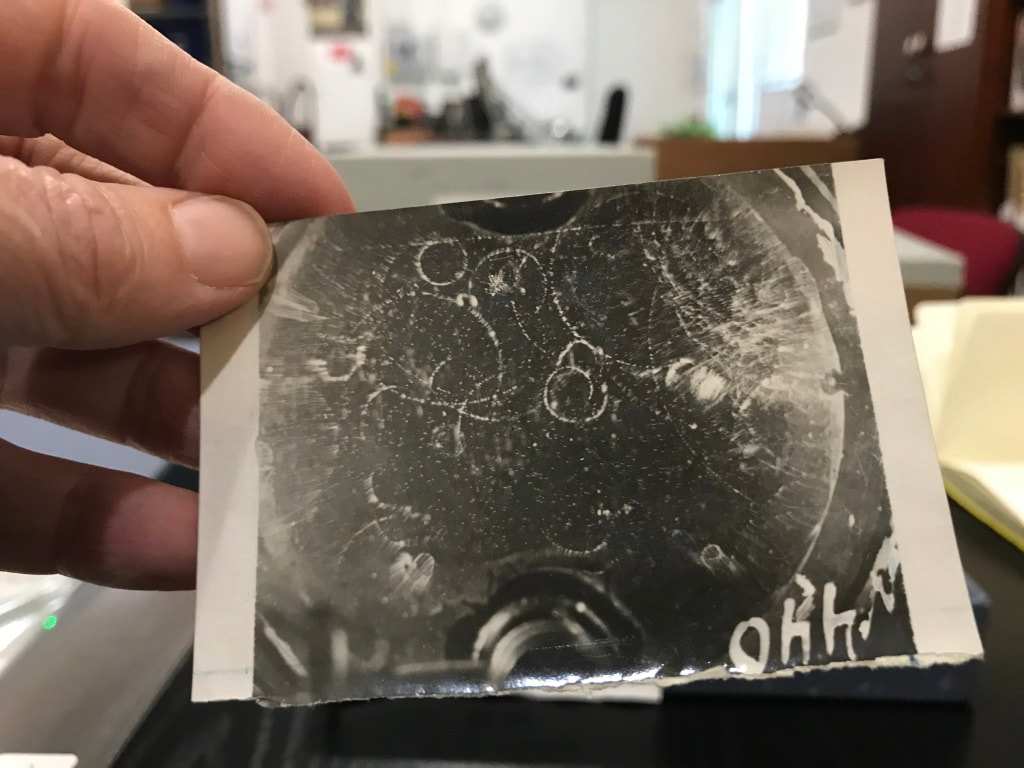
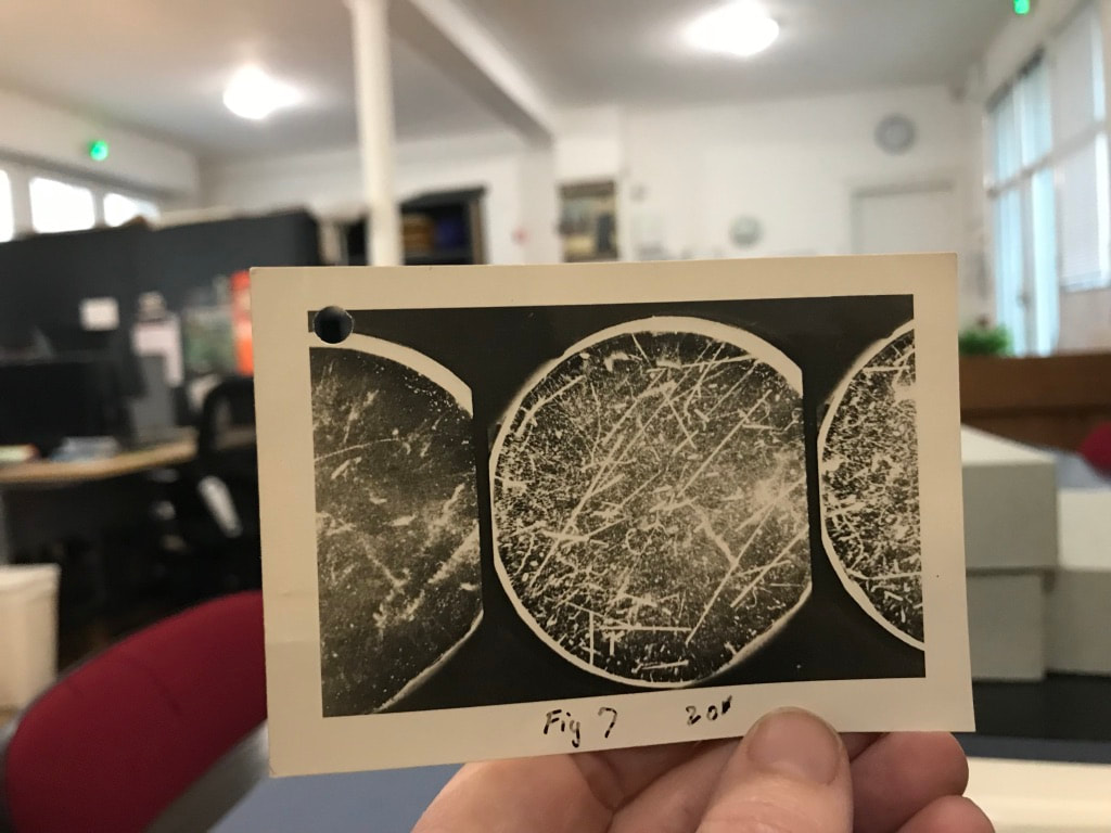
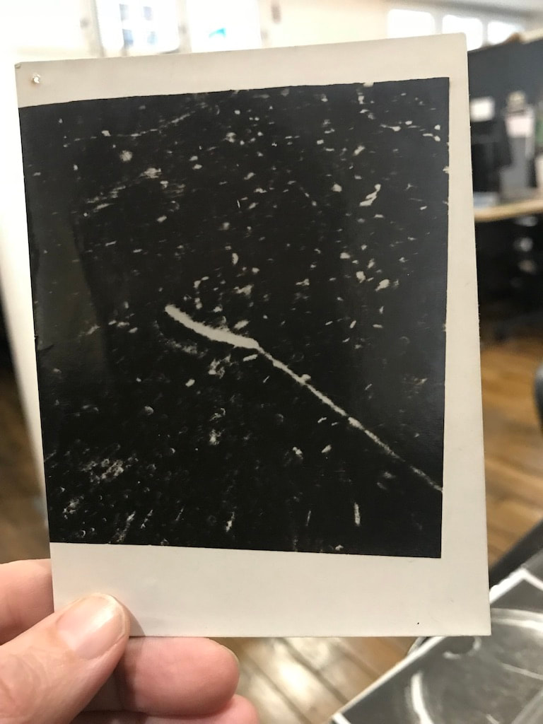
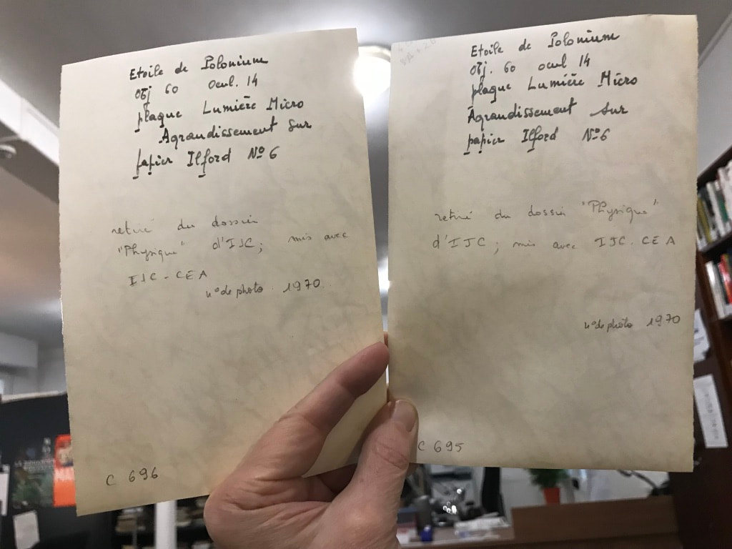
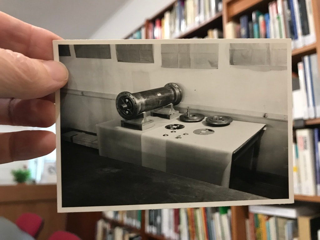
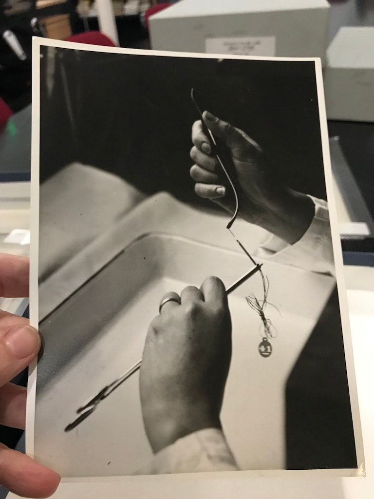
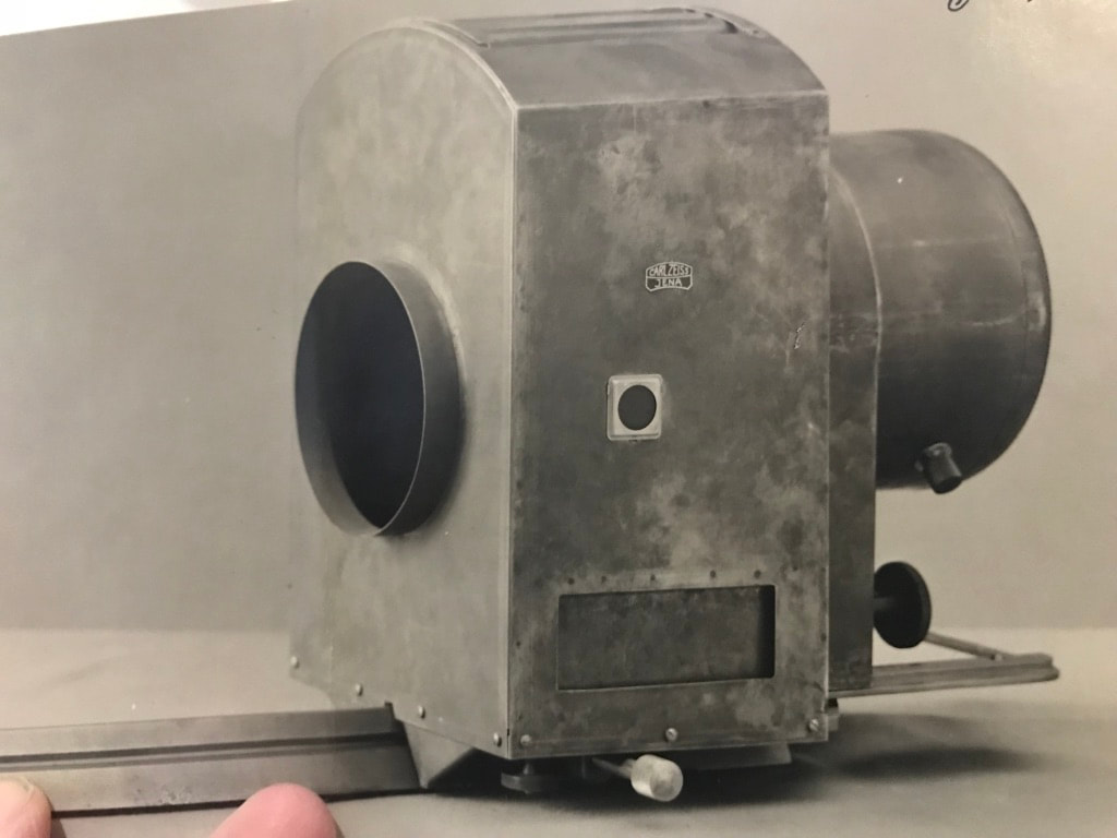
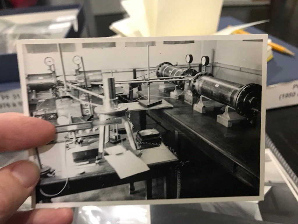
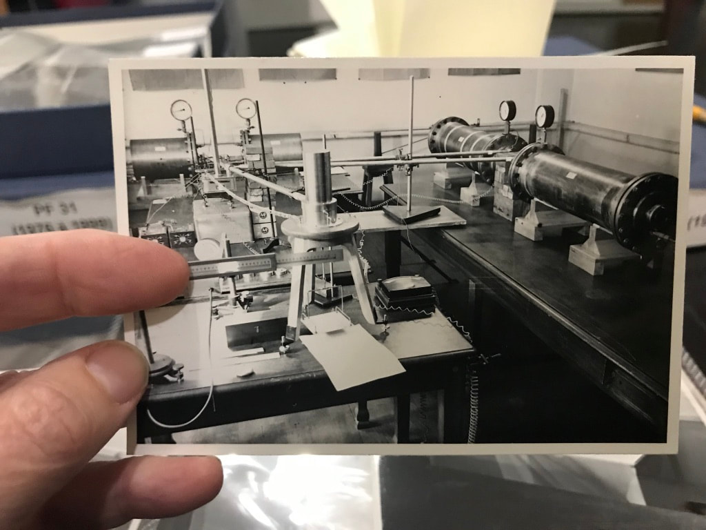
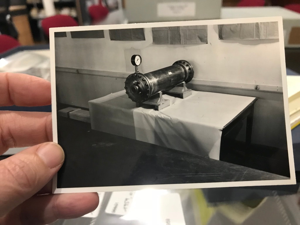
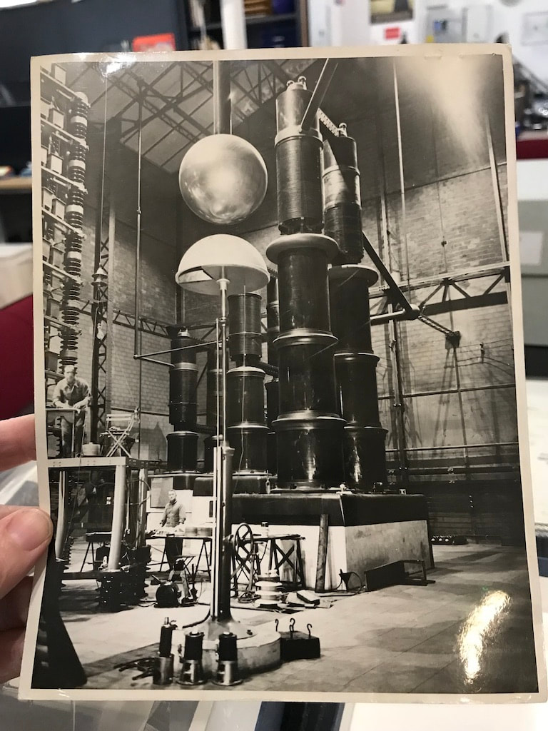
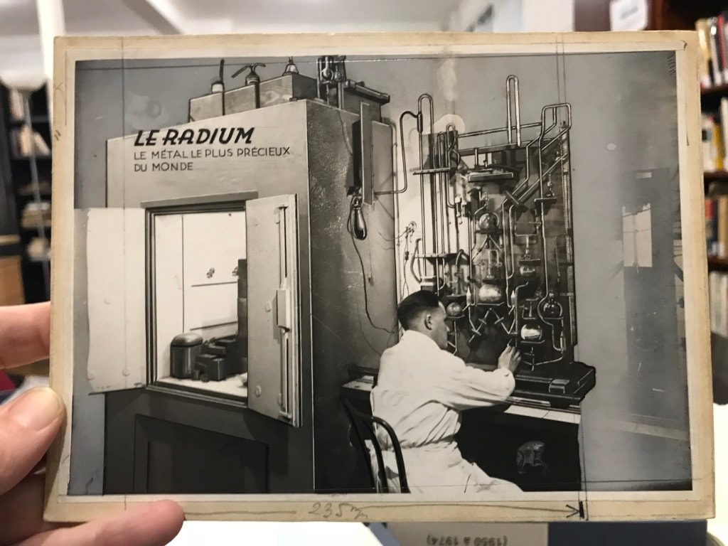
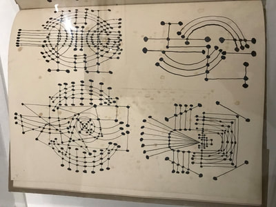
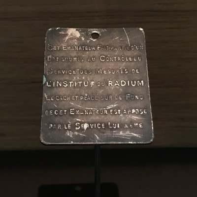
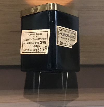
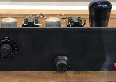
Over the past two weeks, Montse and I have continued to have telephone conversations as well as add to our Dropbox account to develop collaborative preparations. We are planning for her visit to my university in late January, my visit to her university first as preparation in February then in May to work with her students and our exhibition. We are exploring how to structure a pedagogical model for what we will be doing with her class. Stereoscopic imaging and Cyanotype recording continue to be a focus in this initiative. Toward that end, during this time I have used my observation of scientific documentation and scientific equipment at the Curie Museum archives, my visit to multiple contemporary art exhibitions that explore either materials we might be using or processes we might be employing to consider a final format for work produced in the university workshop and classroom. Images attached will give a running narrative of some of my experiences and considerations. Montse
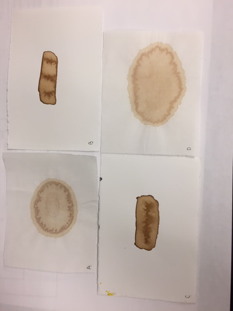
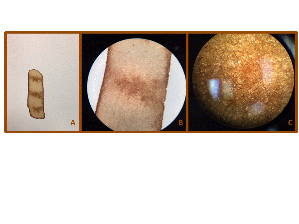
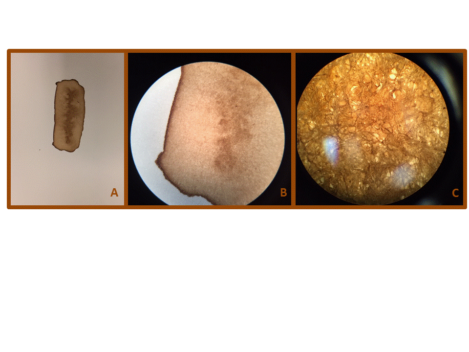
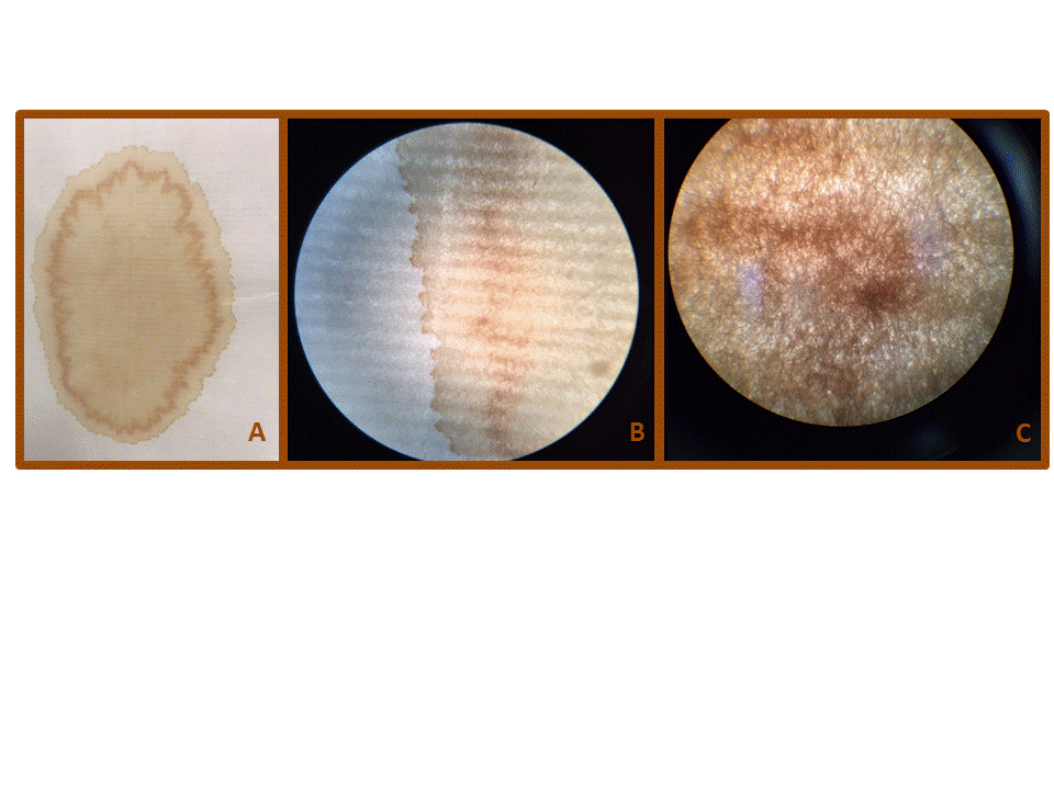
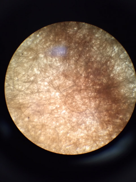
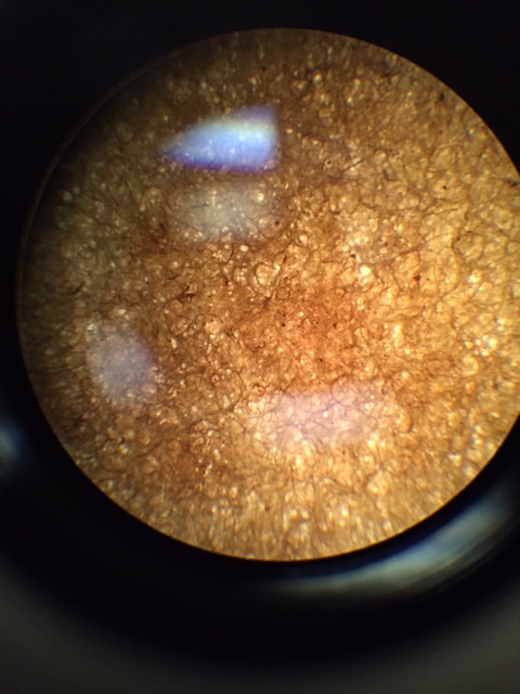
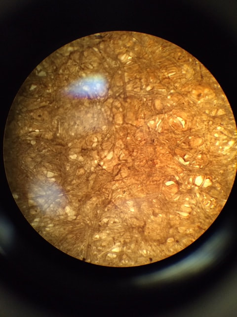
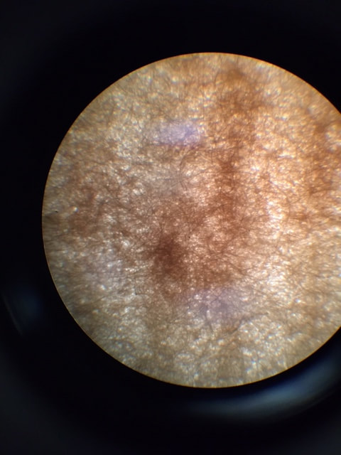
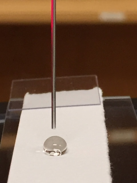
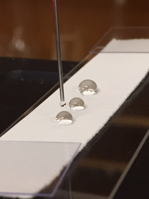
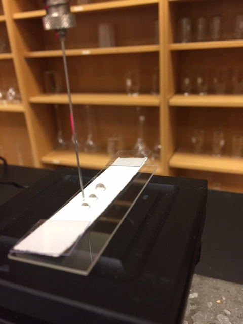
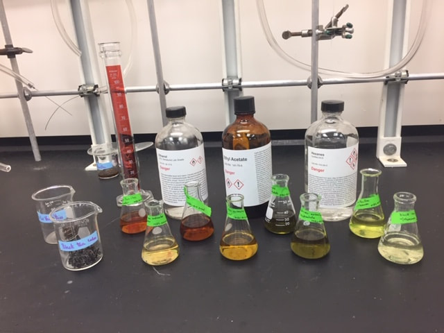
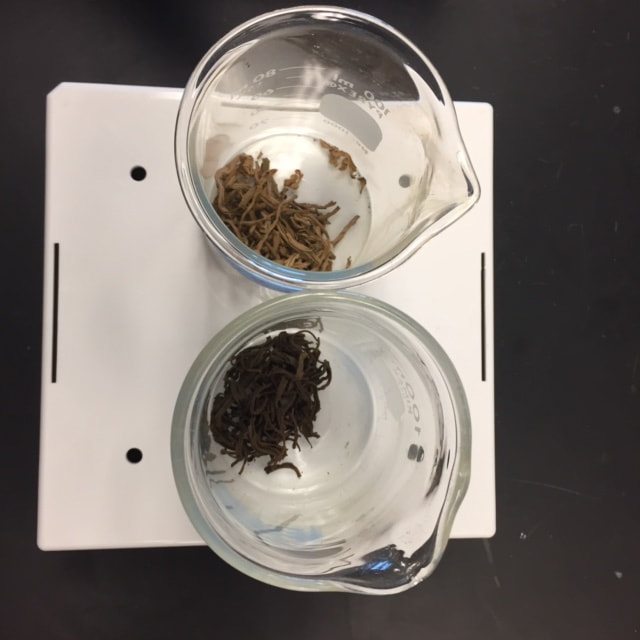
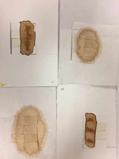
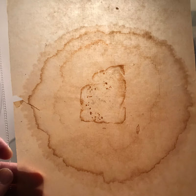
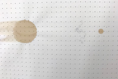
An explanation of the tests were performed the tea samples are as follow. First the samples sent contained two different types of stains; the first group had stains made with tea bags and the second group had stains made with mold. The tea stain samples were further divided into two different groups based on the type of paper that was used to prepare them; one paper was thick and the other was very thin. To perform the tests, four different stain samples were chosen; two with the thick paper and two with the thin paper. Figure 1 shows the four samples used. First, to assess differences in the chemical composition between the two types of papers, a Furier Transform Infrared (FTIR) scan was performed. This technique allows identification of different functional groups, such as hydroxyl substituents, carbonyl substituents, and ether substituents, among others. The results showed that both papers have a similar chemical composition and it is consistent with the reported FTIR spectra of hemicellulose1-2. (Figure 2) Next, the stains were visualized under a microscope using different magnifications. A picture of each one of the samples was taken using a Wolfe dissecting microscope, followed by a picture using a B3 Professional Miotic microscope (Eyepiece WF 10X / 20mm and objective 4X / 0.10 (WD 17mm)). It can be seen that the fibers in the paper from samples 2 and 3 are much thicker and dense compared to the paper from samples 1 and 4. Also, the coloration for samples 2 and 3 is more pronounced compared to samples 1 and 4 (Figures 3-6). An interesting side note is that if one looks at the amplified pictures individually, it is hard to identify them as paper fibers. There can be many interpretations for that picture (See figures 7-10). The next experiment was to determine the contact angle for each one of the paper samples. The contact angle was measured using the Pendant drop experiment and the OneAttension software. The contact angle for the thick paper was 89.87 o ± 0.66 and for the thin paper it was 0 o. This observation was very interesting due to the fact that both materials are the same (which should predict the same contact angle). A possible explanation for this is the fact that the thick paper has fibers that are highly packed, which in turns decreases its porosity. The following pictures (Figure 11-13) were collected while performing the experiment. The next experiment was to extract the tea with different solvents and see if different pigments could be extracted with the intention of isolating different pigments that had different colors (to possibly use them as pigments for a “watercolor that is paint like”). Two different varieties of tea were used for the extraction: the YuLuYanCha and the Laoshan black tea. The solvents used were water, ethanol, ethyl acetate and hexanes. Figure 14 shows the extraction solutions; the front row shows the extractions of Laoshan black tea and the back row shows the extractions of YuLuYanCha. The solvents used for the extraction are in the back row. In order from right to left are: water, ethanol, ethyl acetate and hexanes (most polar to least polar). As it was predicted, various pigments were extracted when solvents with different polarities were used. To check the stability of the color, the solutions were photographed after 72 hours (Figure 15 samples are shown in the same order). The color appears to remain similar to the original test (a further test is coming to check if colors are the same). Figure 16 shows the YuLuYanCha tea after extraction (left) and before extraction (right). A picture of tea and epigallocatechin gallate drawn with those pigments was shown in the previous blog. Also, Jo and I have continued to plan the Sci-Art exhibit. References 1. Célino, A.; Gonçalves, O.; Jacquemin, F.; Fréour, S., Qualitative and quantitative assessment of water sorption in natural fibres using ATR-FTIR spectroscopy. Carbohydrate polymers 2014, 101, 163-170. 2. Ciolacu, D.; Ciolacu, F.; Popa, V. I., Amorphous cellulose—structure and characterization. Cellulose chemistry and technology 2011, 45 (1), 13. Montse
This week Jo and I have been bouncing ideas back and forth about the exhibition/workshop at Kettering University. The list of the items under consideration is:
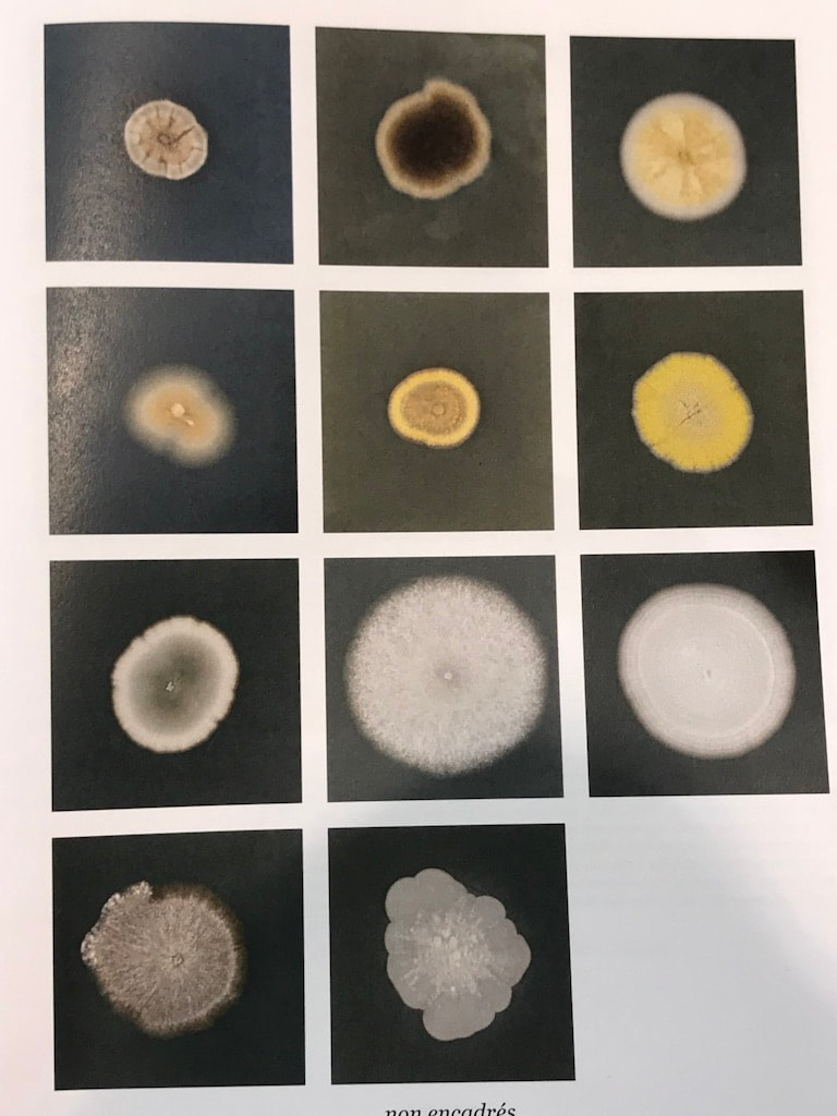
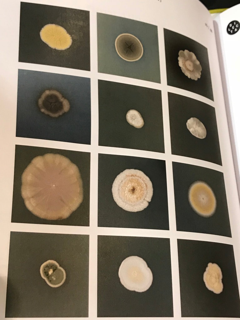
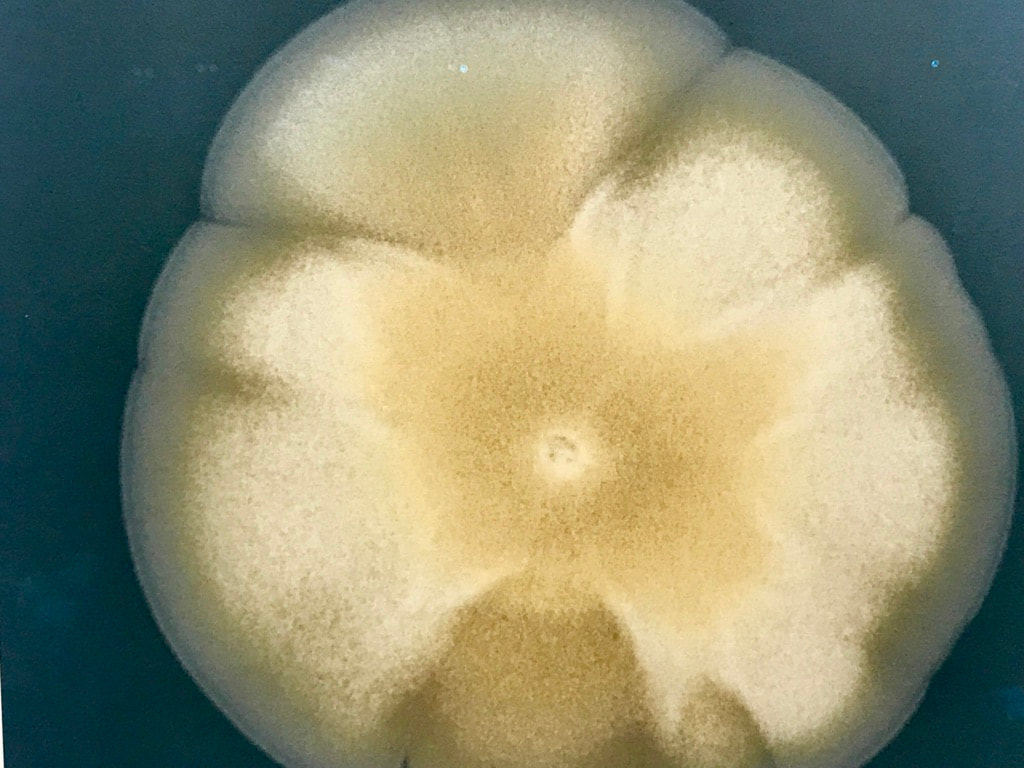
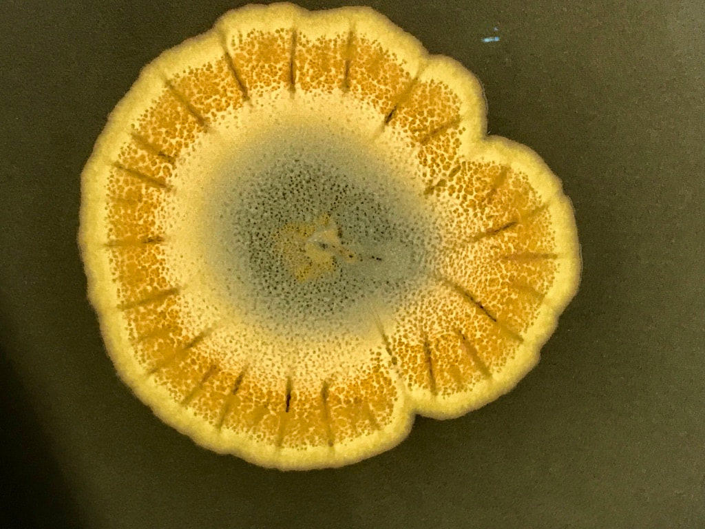
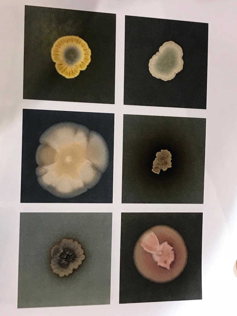
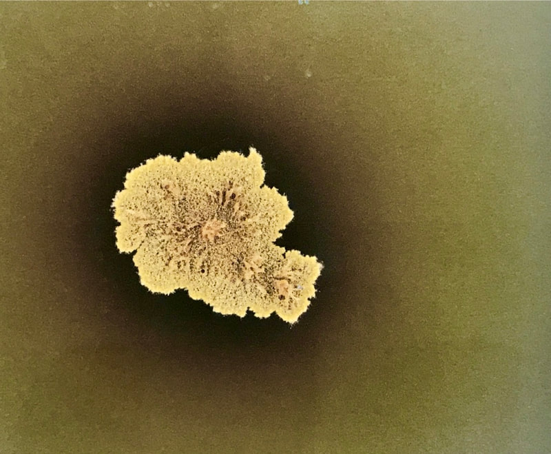
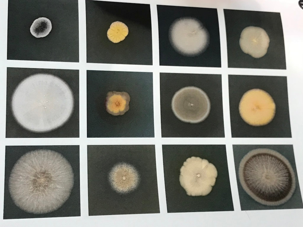
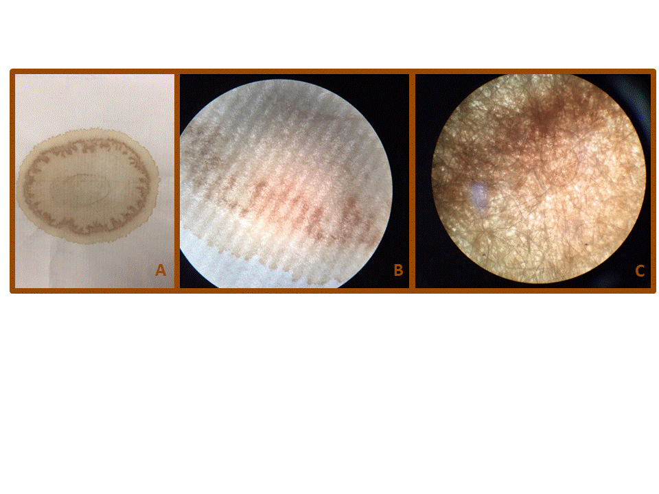
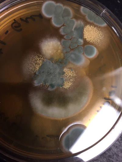
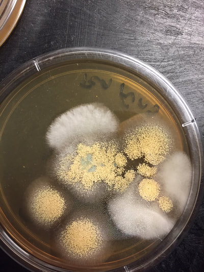
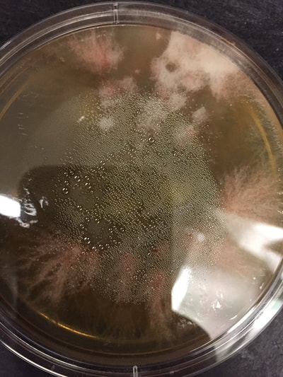
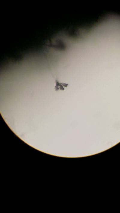
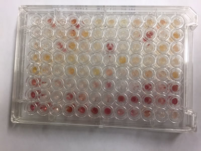
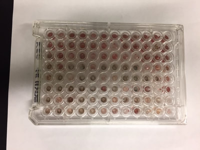
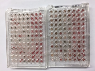
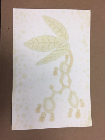
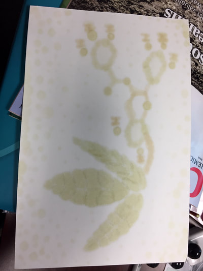
Jo and I would appreciate any input that anyone might have, and we would love to hear others’ ideas or experiences organizing a Sci-Art exhibit/workshop. Montse The mold samples are being tested by students in the Microbiology class. First, the mold was plated (to separate individual types of mold) (Figures 1-3). Pictures of the molds under the microscope are shown in figures 4-6. To identify the molds present in those samples, the FF MicroPlateTM from BIOLOG was used. The FF MicroPlateTM test panel provides a standardized micromethod using 95 biochemical tests to identify / characterize a broad range of fungi including both filamentous and yeast forms.1 Liquid cultures containing individual colonies were grown, and were used in the FF MicroPlateTM, figures 7-9 show the pictures of the 96 well plates. Analysis of all of the data collected to determine the type of fungi is still underway. The experiments performed on the tea stain samples are mostly complete. Each one of the stains was divided into eight sections that were equal in length. Each one of those sections was tested under unique conditions. The reassembled tea samples are shown in figure 10. In conclusion, using the FTIR information, both papers are made of cellulose (see previous blog for details). However, they differ significantly in regard to the thickness and density of the cellulose fibers as well as the surface tension between both papers (see previous blog for details). Another very interesting difference is the apparent variation in hydrophobicity; the thick paper appeared to be a lot more hydrophobic compared to the thin paper. This was an unexpected observation; the reasons for this are unclear and under investigation. Also, Jo and I have continued to think of ideas and activities that can be integrated into an exhibition. One of our ideas is to explore and expand on the use of tea to create something. This idea has been previously explored by other artists; a few examples are shown in the links below (Link 1-5). In order to incorporate the science aspect, I decided to explore a similar idea to Gerard Tonti (link 1), but use the previous extracts (see previous blog for details) to create a watercolor paint. To compare how the tea paint would look versus the extract paint, I composed two drafts: one using real watercolors (figure 11) and one using extracts (figure 12 front of paper figure 13 back of paper). In both, I attempted to draw the tea leaves and one of the most important antioxidants in tea: epigallocatechin gallate. Links: Link 1 https://www.dailymail.co.uk/news/article-2452341/Putting-TEA-art-Artist-swaps-paint-hot-drinks-create-intricate-portraits.html Link 2 https://www.huffingtonpost.com/2015/02/05/tea-bag-portrait-art-red-hong-yi_n_6622180.html Link 3 http://redhongyi.com/portfolio/untitled-tiger-with-tea-leaves/ Link 4 https://mymodernmet.com/miniature-paintings-tea-bags-ruby-silvious/ Link 5 https://www.brandsouthafrica.com/play-your-part-category/play-your-part-news/women-recycle-tea-bags-to-make-art 1. www.biolog.com, FF MicroPlateTM Manual. Jo
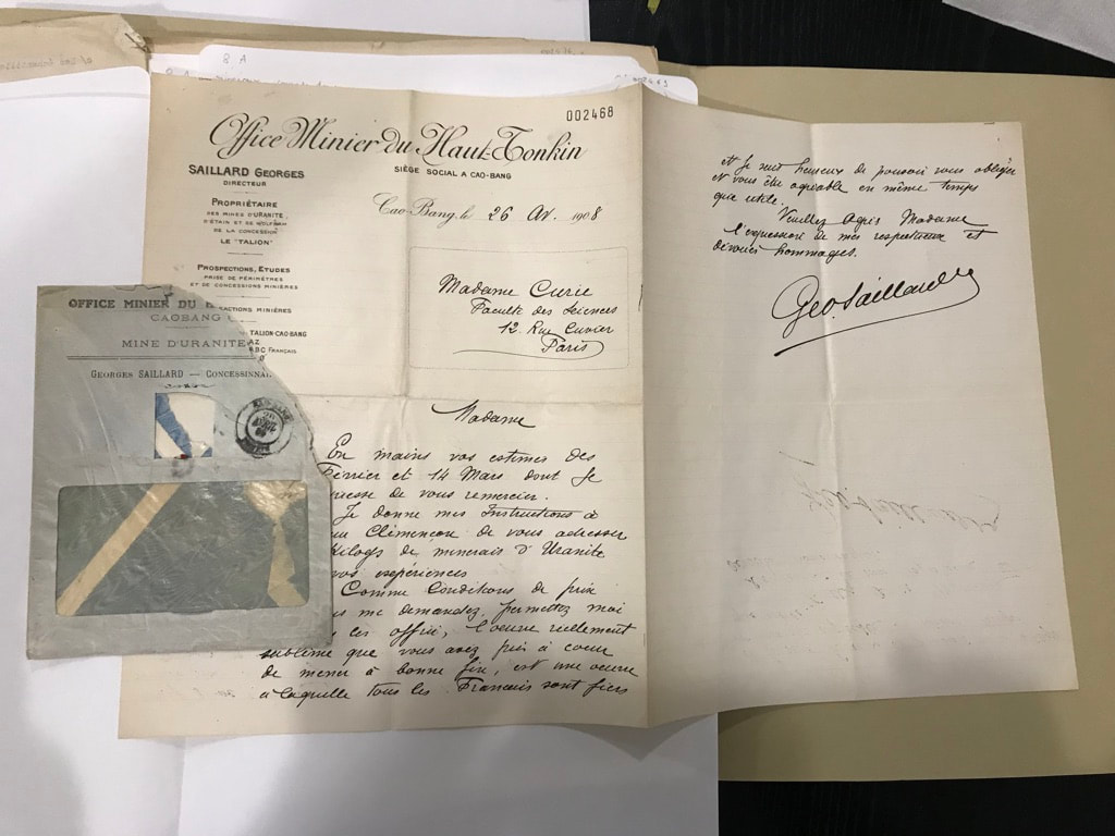
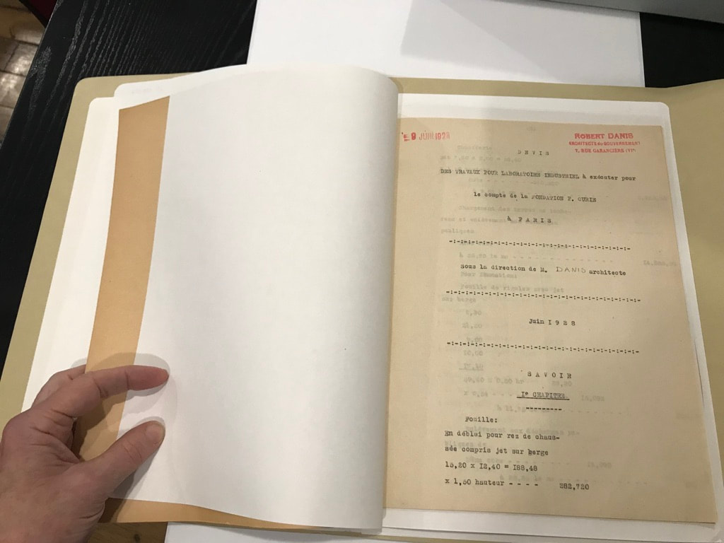
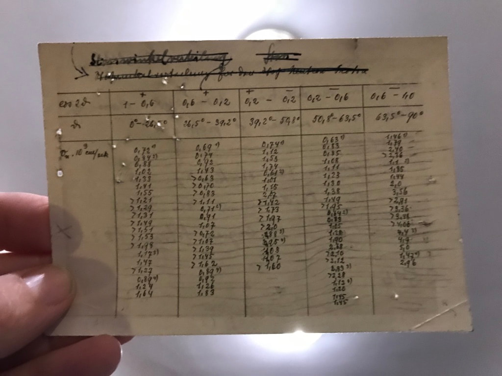
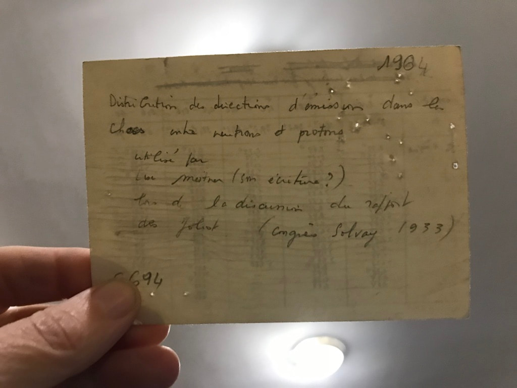
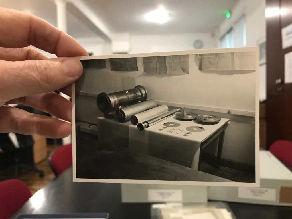
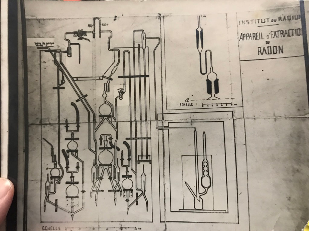
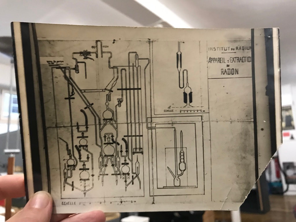
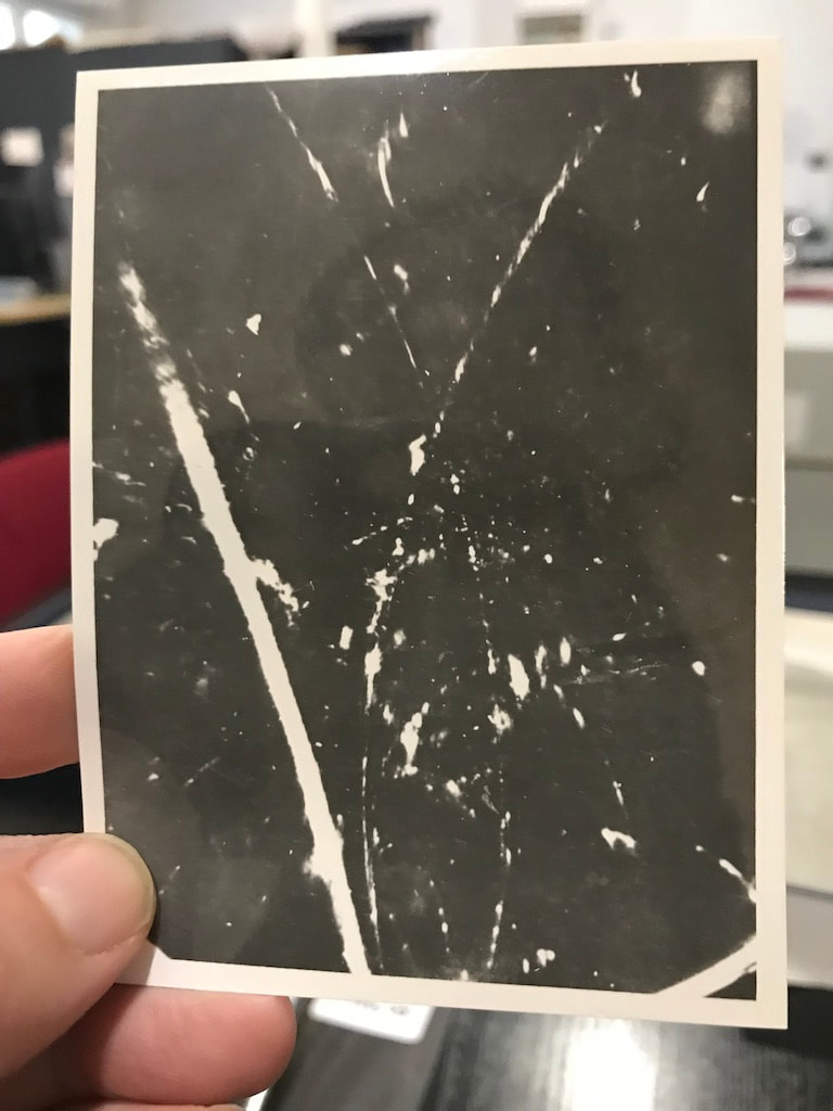
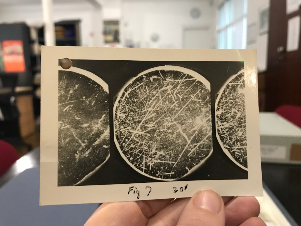
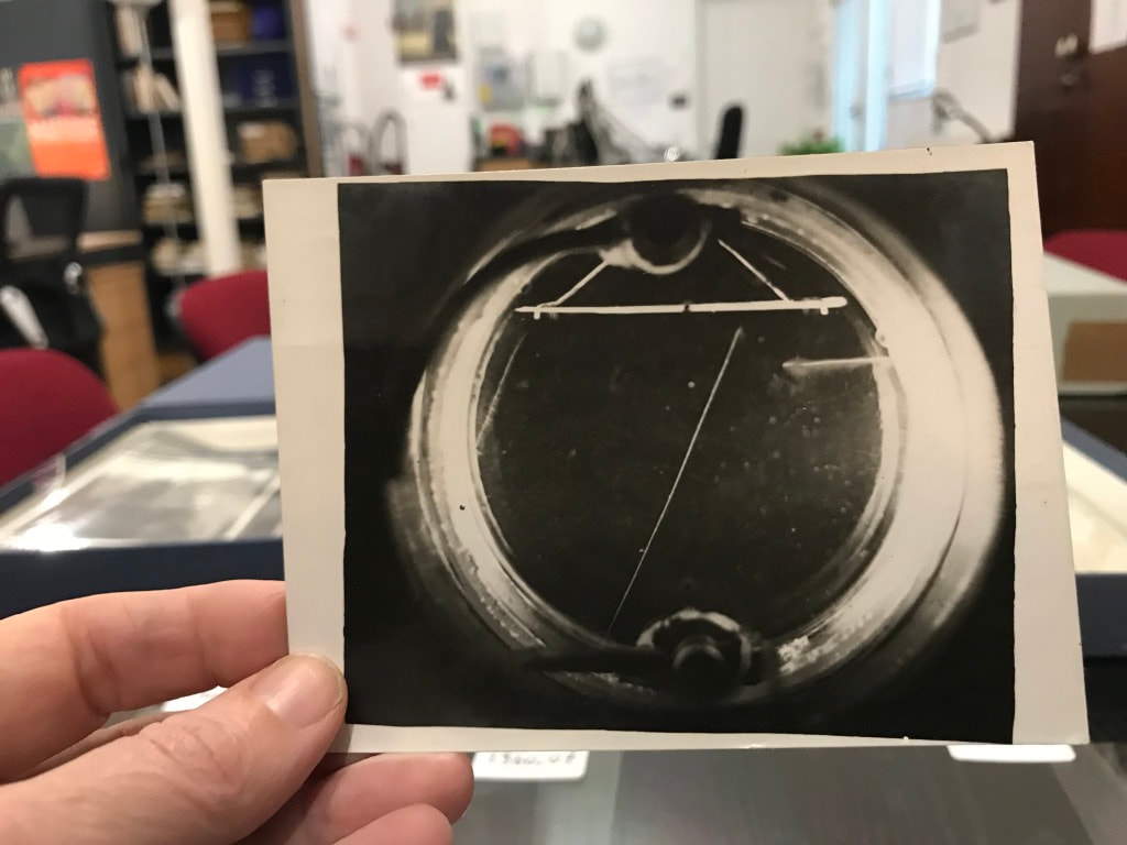
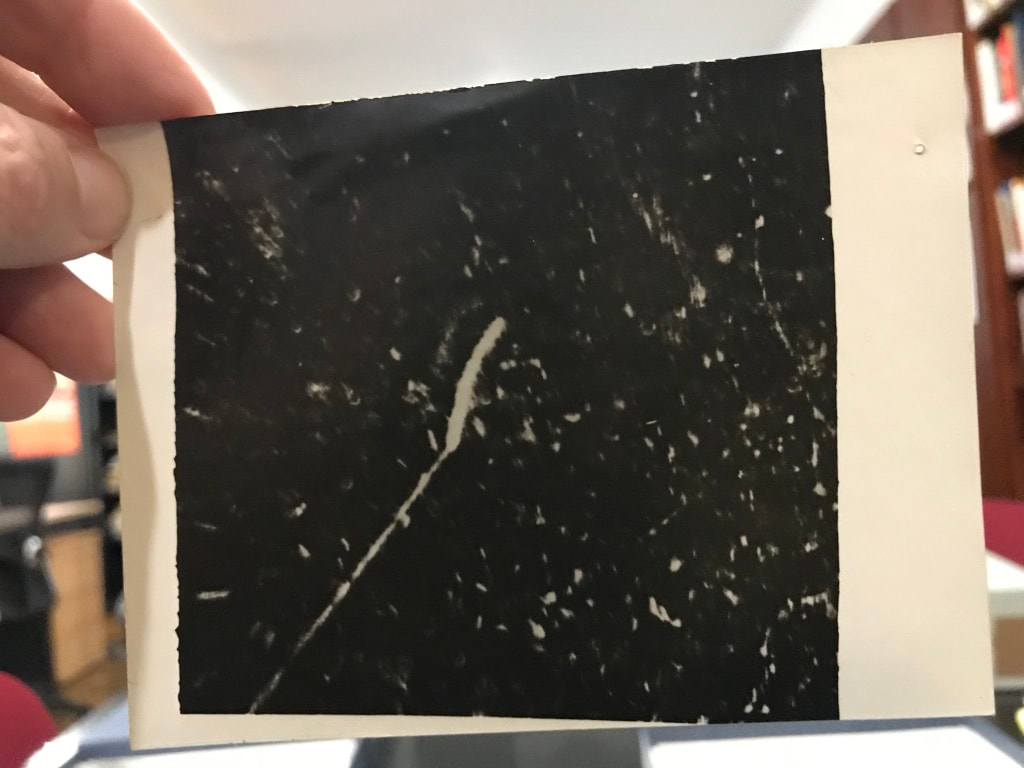
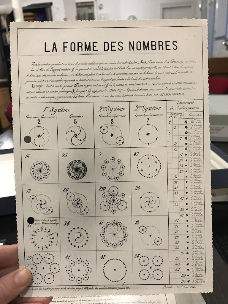
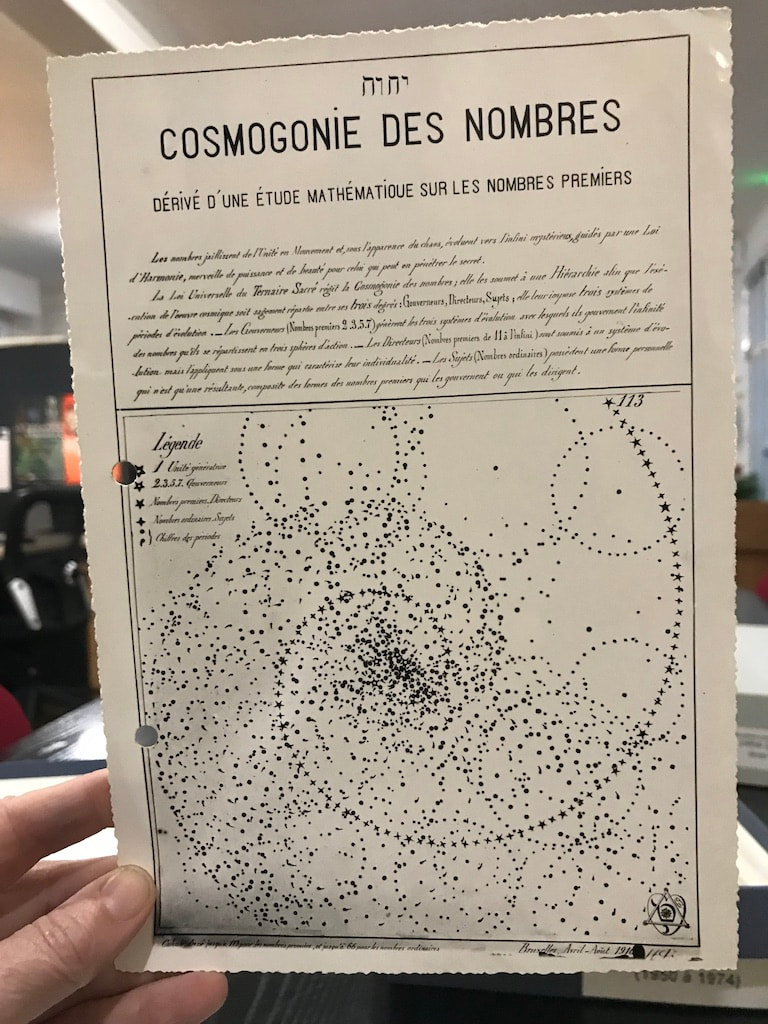
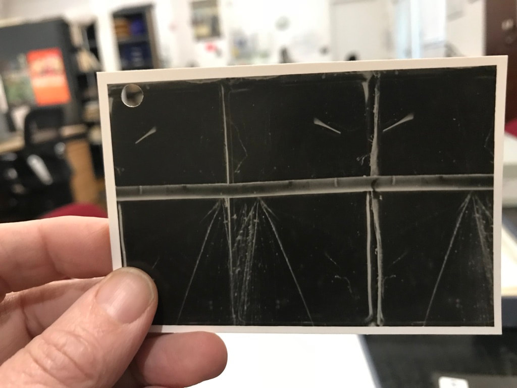
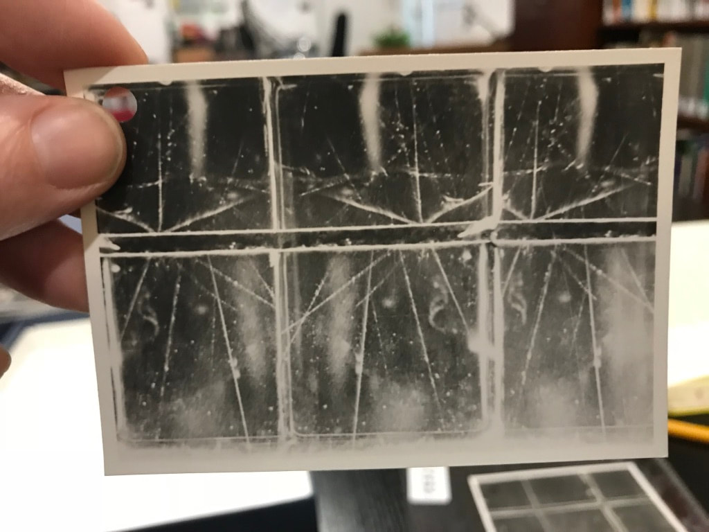
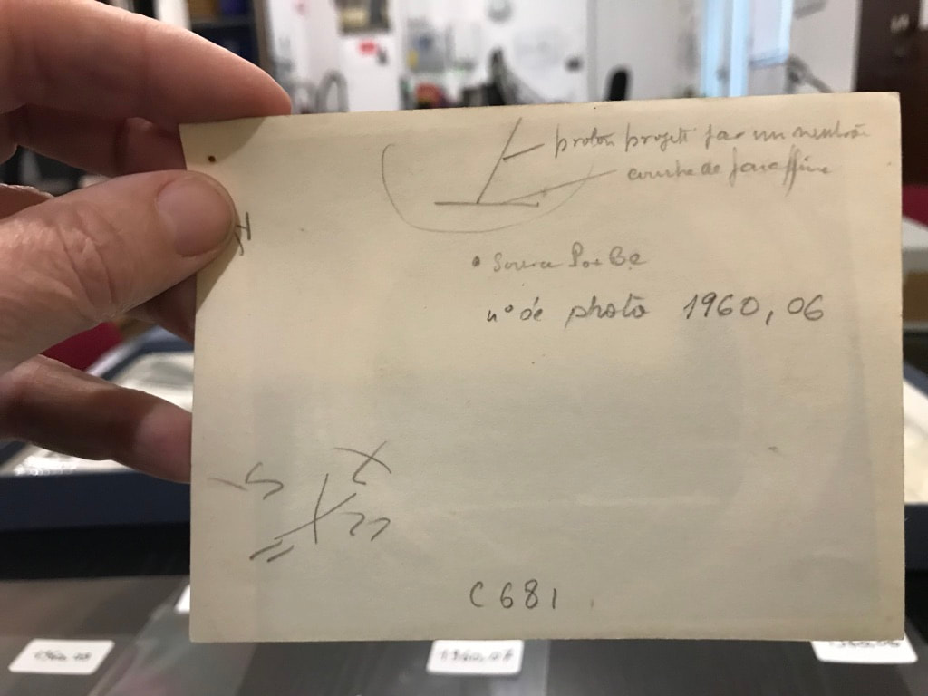
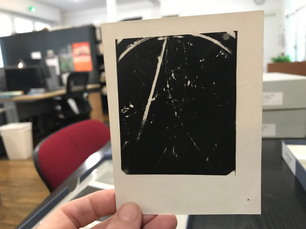
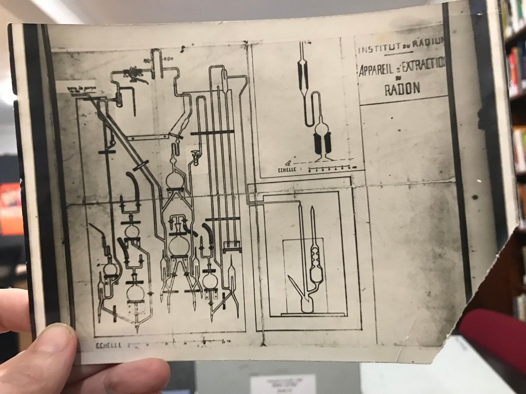
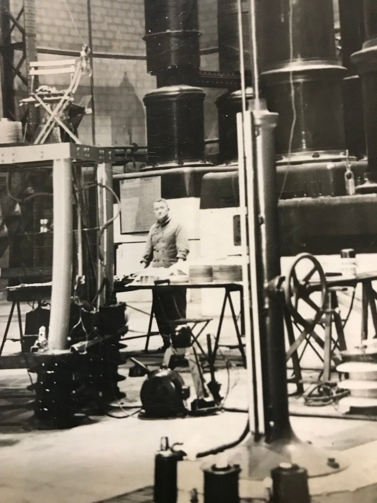
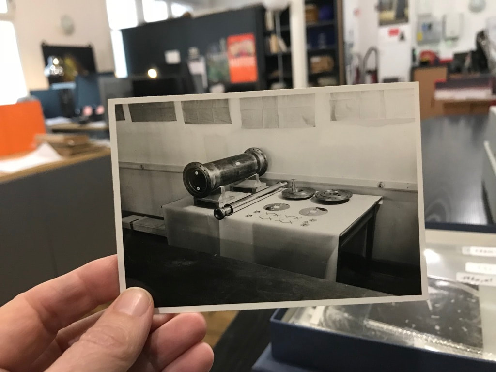
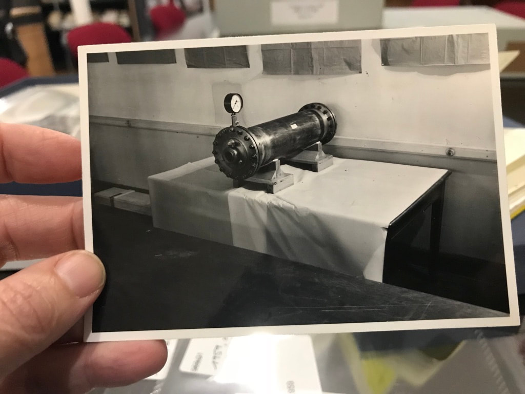
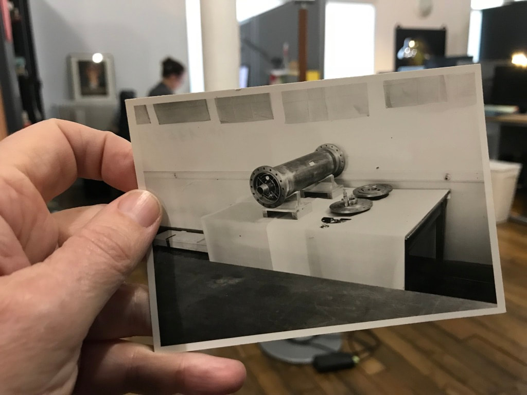
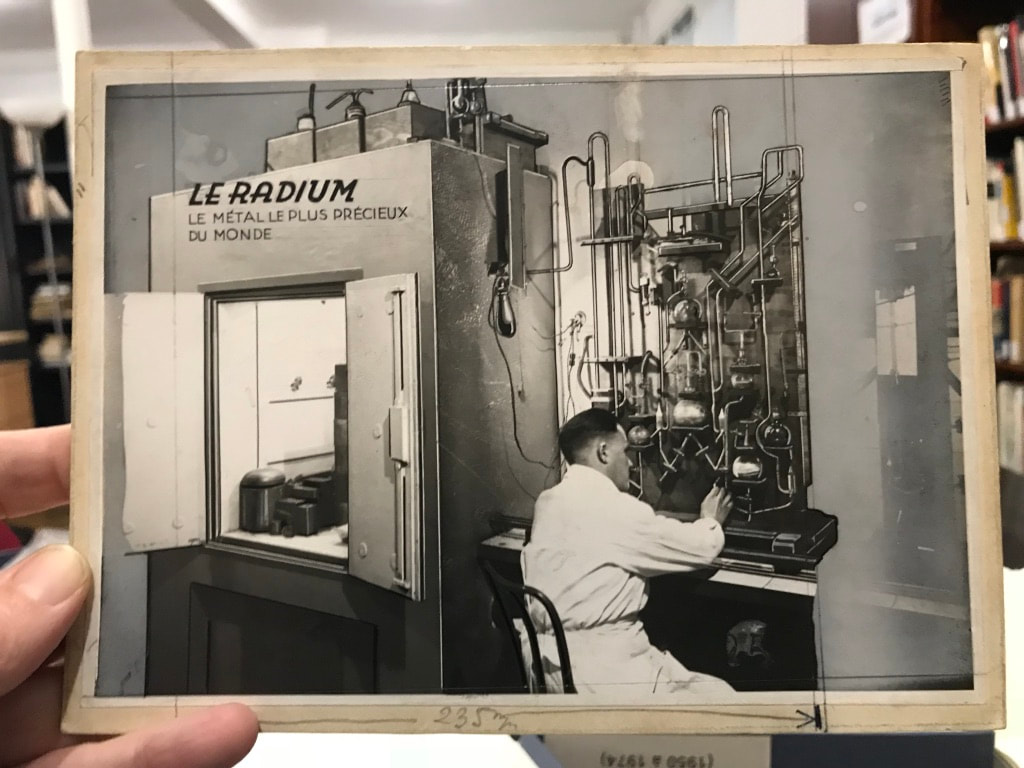
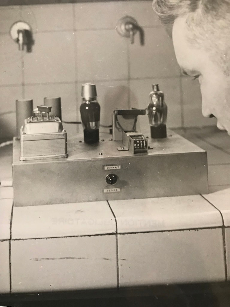
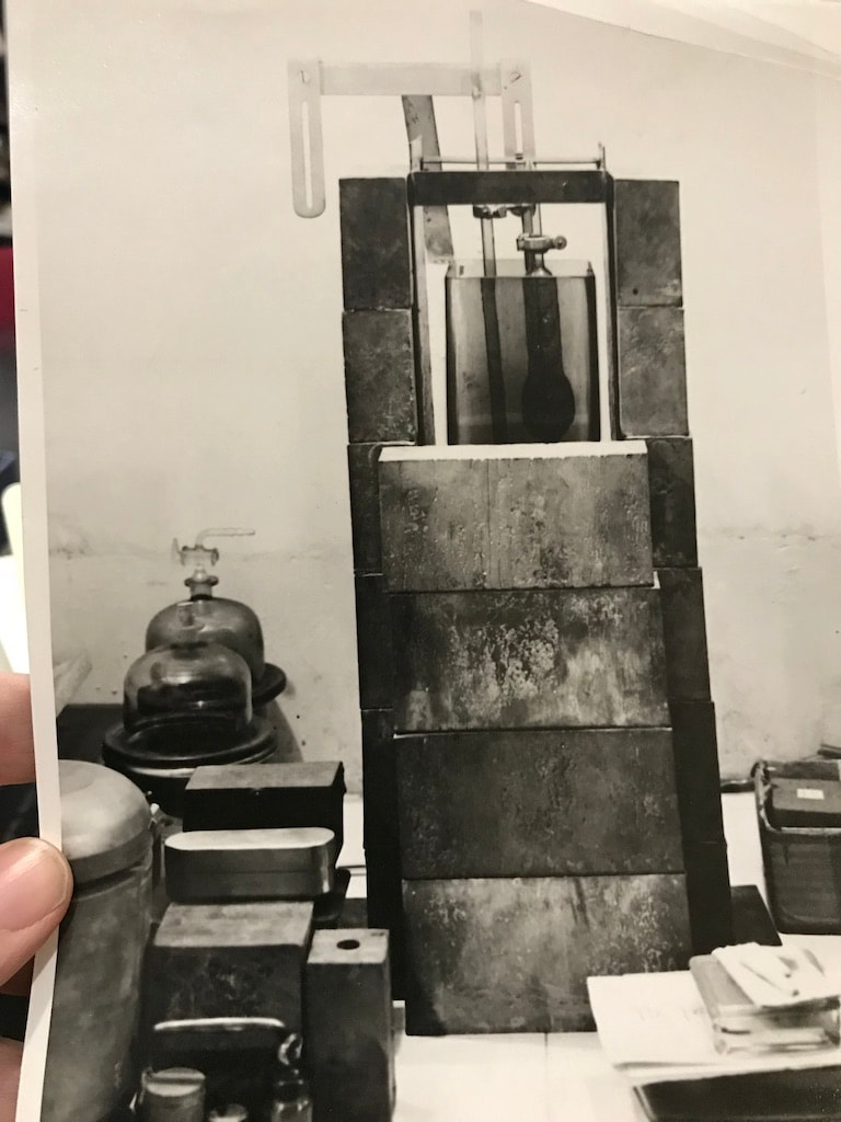
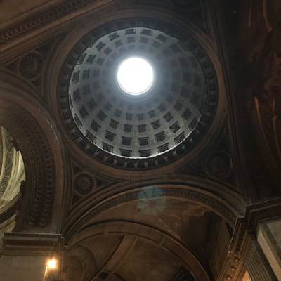
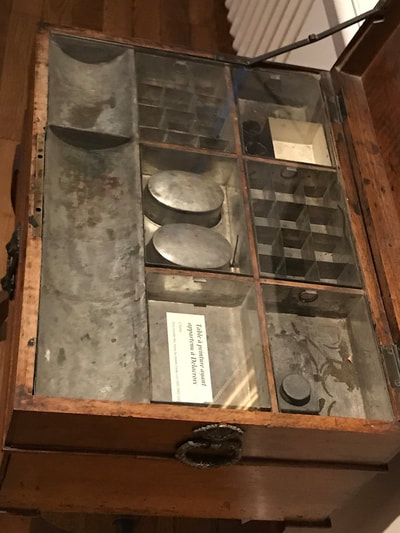
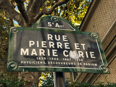
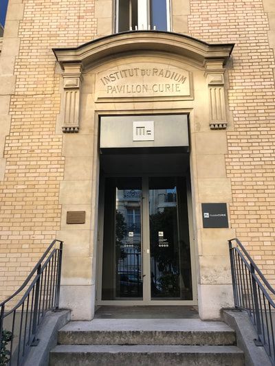
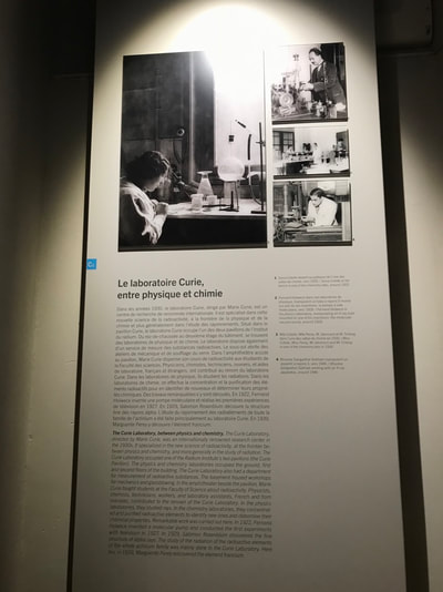
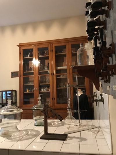
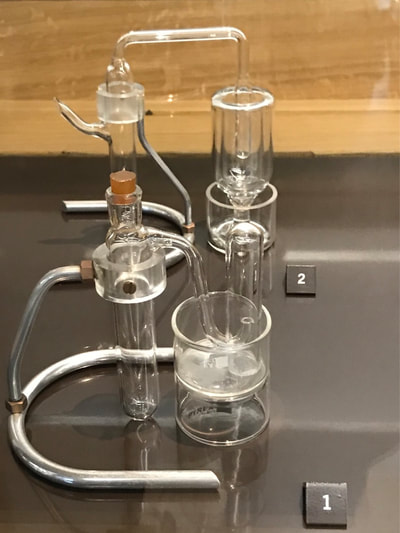
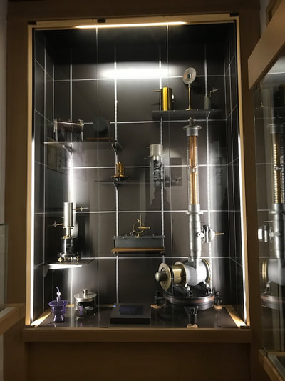
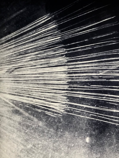
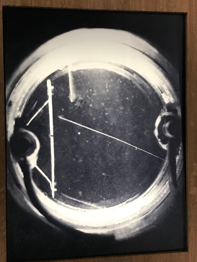
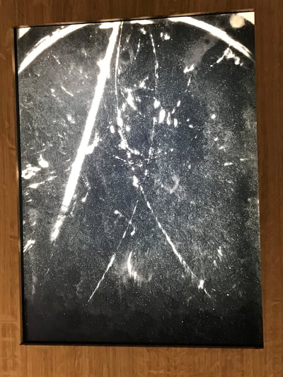
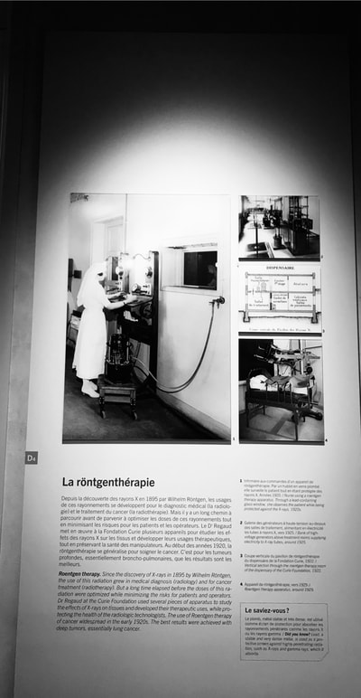
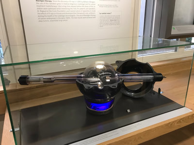
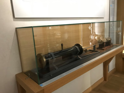
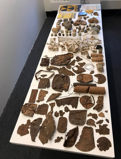
As mentioned in my last entry, this week I was visually researching/getting inspiration from visiting in Paris with other artists and curators a number of art and science museums as well as searching out obscure cultural locations and events. I have a number of examples of locations and works which have inspired me and have labeled and arranged them to create a kind of spontaneous and chance-based narrative (a type of artform, perhaps). Also following is a copy of my email communication with the Curie Museum staff/curators as I first visit and then try to gain entry to the collections, to photograph with a stereoscopic camera the Curie lab equipment and notebook pages: Dear Professor Yarrington, First of all, thank you for your interest in our collections ! Before giving you an answer I need to discuss your request with the head of the historical resources department, who will only be available next week. I will contact you as soon as I have more information, sorry for the delay. Best regards, Aurélie LEMOINE Aurélie LEMOINE Archiviste Musée Curie - UMS 6425 CNRS/Institut Curie adresse postale : 11 rue Pierre et Marie Curie 75248 Paris Cedex 05 adresse de consultation archives : 21 rue Tournefort 75005 Paris Tél. :+33 (0)1.56.24.55.49 [email protected] Rejoignez-nous sur Facebook De : Yarrington, Kathryn J. <[email protected]> Envoyé : lundi 5 novembre 2018 15:11:38 À : Klapisz Adrien Cc : [email protected]; documentation.musee Objet : Marie Curie archives Dear Adrien, It was wonderful meeting you last week and I thank you for your kindness in spending so much time at the end of the day to help me with my visual art research and process focused on Marie and Pierre Curie’s discoveries. As mentioned to you when at the Museum, I am in Paris for 5 weeks from late October to December 2018 to research and photograph at the National Library and at your museum, specifically focusing on Madame Curie and her journals.. By way of an introduction to your other colleagues at the Museum, my name is Jo Yarrington and I am a visual artist and full professor at Fairfield University with a primary focus in printmaking and photography, although I also have been developing work in comprehensive installations which have included glass production and artist book series. My website is www.joyarrington.com>. I live in New York City and have been awarded a yearlong sabbatical leave, 2018-2019, to continue my focus on uranium as product, process and political “code”. I have two solo exhibitions coming up in spring 2019 in museums in New Jersey and China, both using uranium as product and metaphoric lens. Because my work is multi-disciplinary, this focus has taken me from documenting Nuclear Power Plants in the United State and Europe to more recently photographing with a small group of collaborators abandoned uranium mine sites in the state of Utah and the Navajo Nation (located in southern Utah) where we have been researching the current politics and local history (including physical artifacts found at the mines). Our photographic work also extends into alternative photographic processes and we have been using uranium to make uranium prints (a process my colleague, photographer Morgan Post, recently resurrected from a mid-1800’s formula). Using this formula, we employ either negatives from images taken at the mines or, specifically in my case, as photograms derived from objects, a la Man Ray. While in Utah, among other mines, I visited the Temple Mountain Mining Complex from which uranium was extracted and shipped in the late 1800’s to France to be used by Madame Curie for her experiments with pitch blend extraction. As mentioned, I would be interested in the following help. My aim is to photograph laboratory objects and manuscript pages not yet digitalized. For this, I am using a vintage stereoscopic film camera (commonly known as a Viewmaster) to photograph the Curie notebook pages and laboratory objects, thus creating an optical dimensionality. I will have an example of one of the Viewmaster cards and a Viewmaster to examine with me, to aid in clarification. Famed photographer Steven Shore also used this method in some of the groundbreaking series he did in the 1970’s. Steroscopic photography relates to the historic time in which the notebooks were written by Marie Curie and provides a metaphoric resonance, a way to look at scientific discoveries, as Marcel Proust would say, through “two lenses”. Although digitalized photographic reproductions probably are available of most items, it would compromise the integrity of that central idea for me to not be able to photograph the existing pages and/or objects, as outlined in the following. You mentioned that I should be specific since different curators are responsible for each area in which I wish to view and photograph: * Notebook pages: To view and subsequently photograph non-digitalized Curie notebook pages currently in the archives * Scientific images: To view and subsequently photograph the science-based images such as the photogaphs of the Wilson cloud chamber, the trajectory of alpha rays emitted by a source of radium, trajectories of positive and negative electrons and any other derived image in which special equipment was used to explore reactions and emissions. * Glass-based laboratory equipment: To view and subsequently photograph glass laboratory equipment, especially that which was used to extract radium or was used as a health-based light emission (such as the black light tubes). I would be most grateful for any help you can offer to move forward with my research. I am available this coming Wednesday and Thursday (8, 9, 10 November) to visit the Museum again and perhaps discuss with each curator what I might be able to see and what I might, at a later date, be able to photograph. I look forward to hearing from you at your earliest convenience. Sincerely yours, Jo Yarrington Professor of Visual Art Fairfield University United States From: Klapisz Adrien <[email protected]> Date: Friday, November 2, 2018 at 4:48 PM To: Kathryn Yarrington <[email protected]> Cc: "[email protected]" <[email protected]>, "documentation.musee" <[email protected]> Subject: [External] Marie Curie archives Musée Curie : http://www.calames.abes.fr/pub/curie.aspx#details?id=FileId-480 http://www.calames.abes.fr/pub/curie.aspx#details?id=FileId-450 National Librairy : Bnf https://archivesetmanuscrits.bnf.fr/ark:/12148/cc7376q https://gallica.bnf.fr/services/engine/search/sru?operation=searchRetrieve&version=1.2&query=%28dc.title%20all%20%22Pierre%20et%20Marie%20Curie%22%29&keywords=Pierre%20et%20Marie%20Curie&suggest=1 archives et informations : "documentation.musee" <[email protected]> Jo
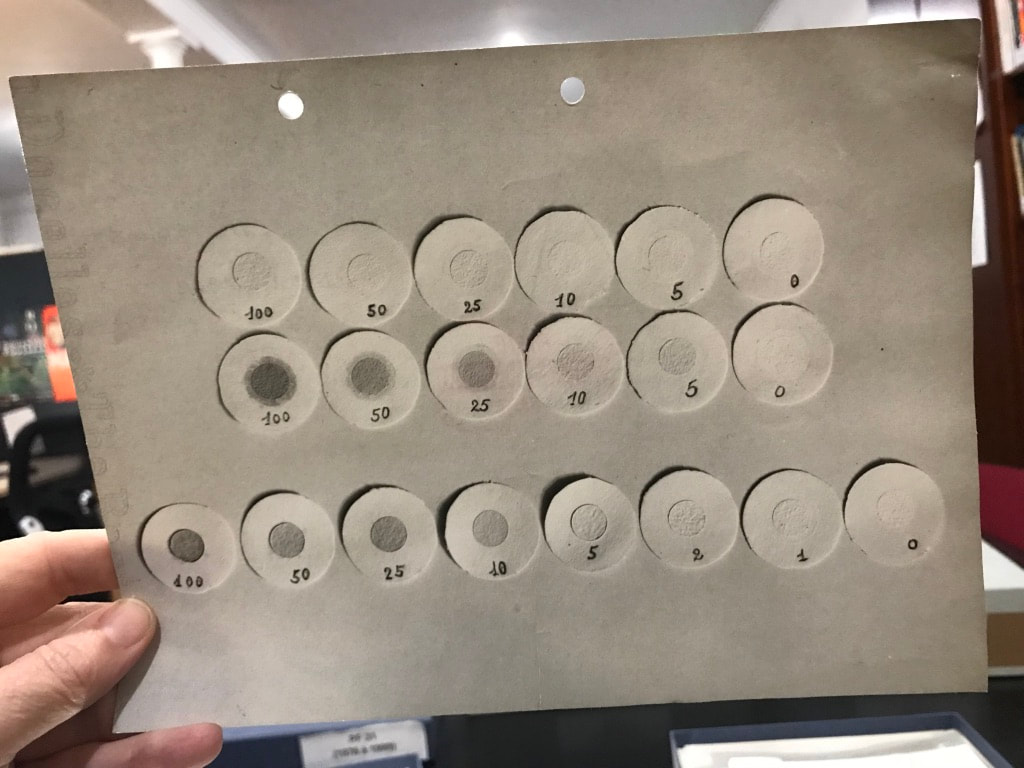
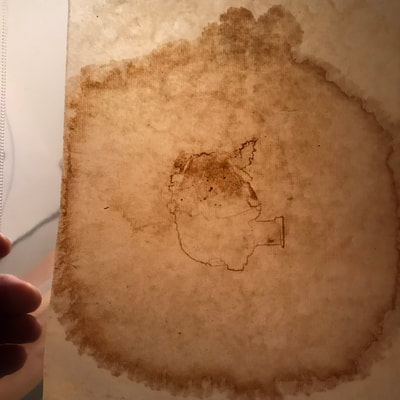
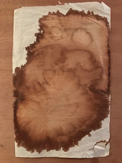
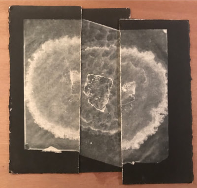
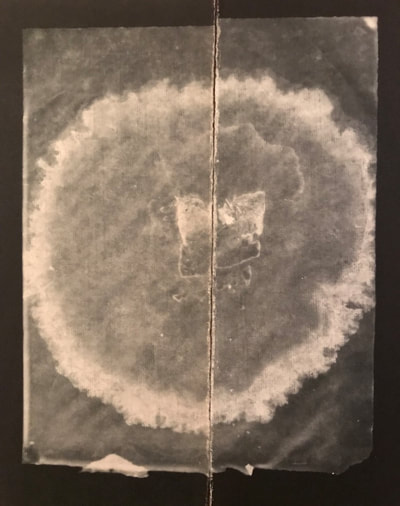
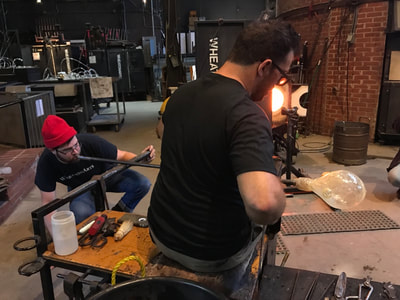
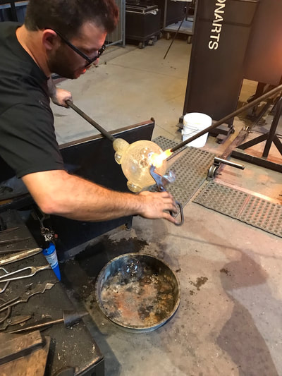
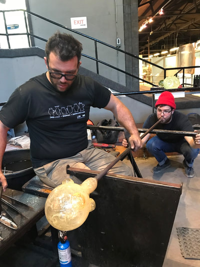
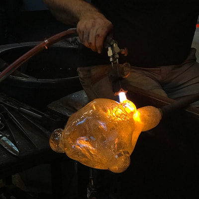
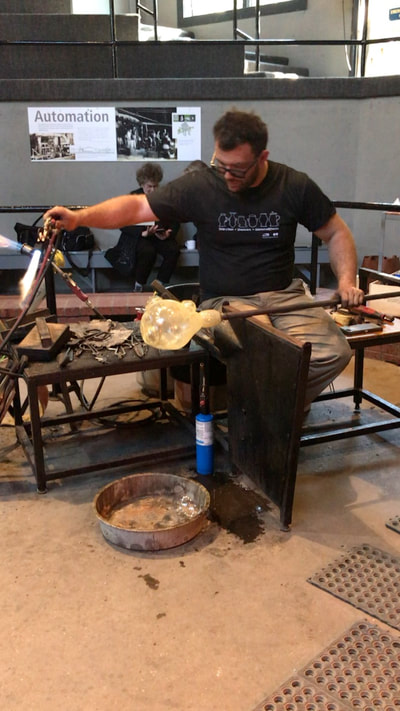
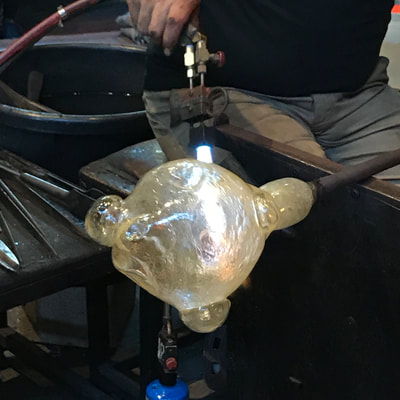
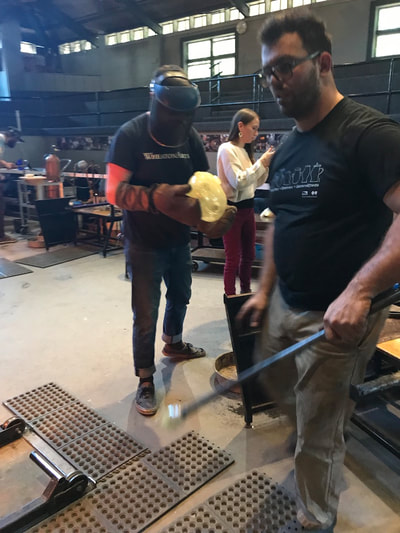
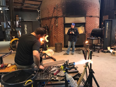
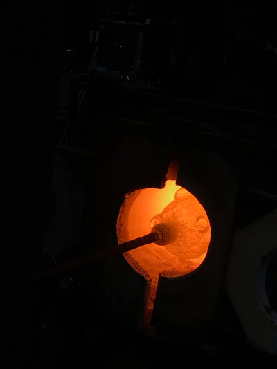
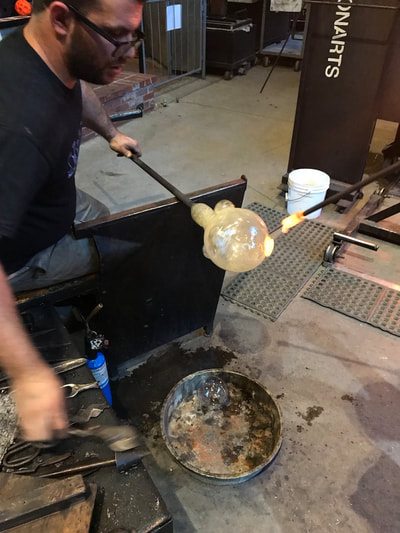
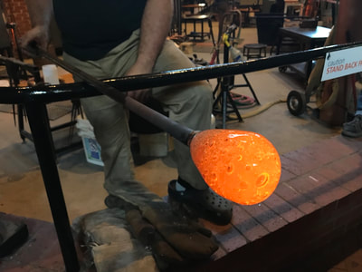
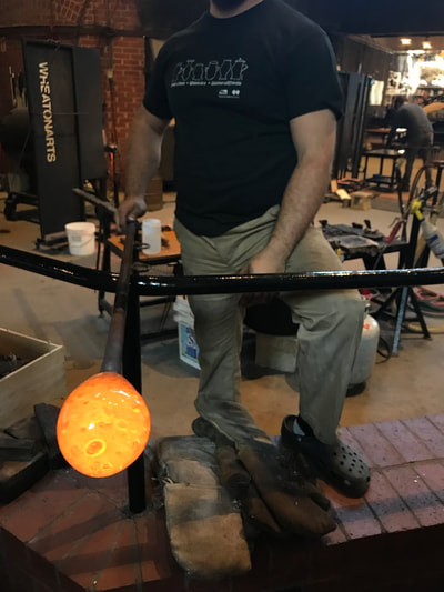
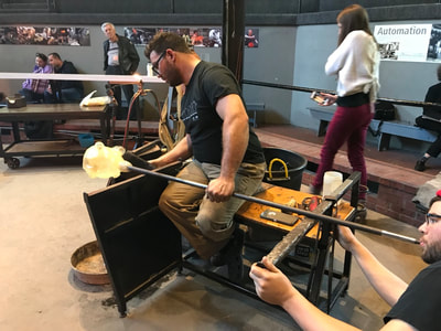
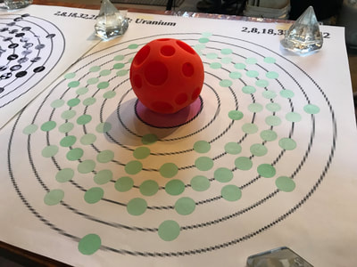
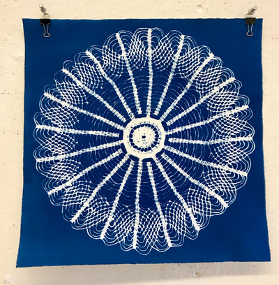
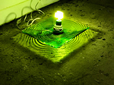
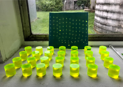
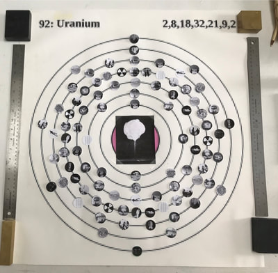
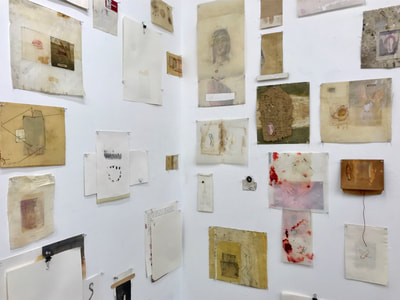
As Montse and I begin to structure an interdisciplinary visual collaboration from workshops that originate in her lab, to working on research with her students, to presenting a visual workshop to developing an exhibition at her university in the spring, its interesting to see the emerging visuals from her digital documentation of microscopic samples. When I return from Paris on December 1, I hope to make negatives from these images and use them for the Cyanotype process. We also are continuing to discuss Cyanotype chemistry using iron-based materials she is using in her research. Last week I included examples of some of the tea stains I created on paper and then waxed during a residency in the early 1990s, but did not label or number them so am resubmitting to show materials used. Also included in the visuals are this week’s formation of more uranium glass pieces at the Wheaton Arts and Cultural Center with gaffer Skitch Manion. He is helping me to create Uranium Games, a political “game” based on the atomic configuration of the element U. As I mentioned to Montse, we also could use the digital images from the microscope slides for the glass technique shown using a photographic decal fuse. This week I am visually researching/getting inspiration from visiting in Paris with other artists and curators currently in the city a number of art and science museums as well as searching out obscure cultural locations and events. I should have a number of examples of locations and works which have inspired me and will post for the next blog. Montse
This week Jo and I have been talking about having an exhibition/workshop together at Kettering University. At this point we have been brainstorming and we came up with the following topics to explore:
Currently, we are thinking of using the following ideas to move forward:
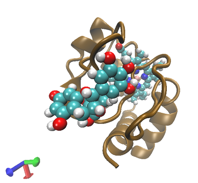
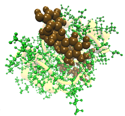
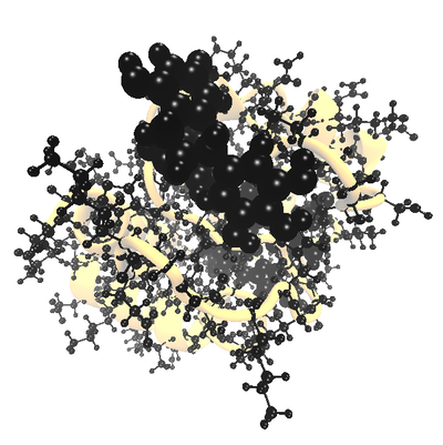
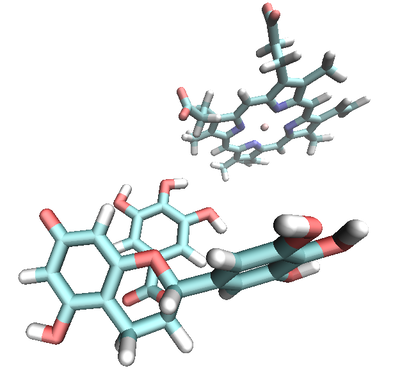
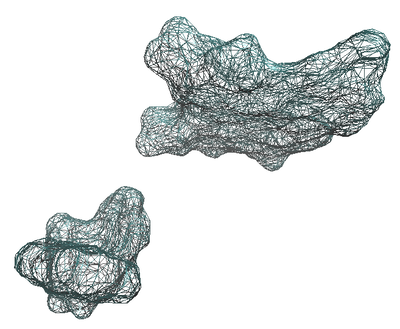
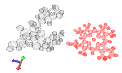
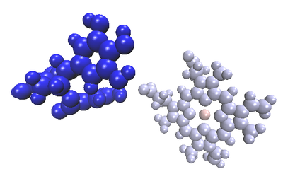
Montse This last week I have been reflecting on how I can take this experience and share with others, especially my students. I have been thinking about how I could incorporate Cyanotypes into my laboratory class, but have come up with nothing concrete yet. Last week, I went back to previous preliminary work I have done. In this work I used SwissDock (Link 1) to simulate the molecular interactions between bovine cytochrome c, (2B4Z1), a protein involved in the respiration process) and epigallocatechin gallate (EGCG), one of the major components in tea). The program provides all of the possible interactions between these two molecules. In the past, when I analyzed the modeling results, I was careful to pay close attention to the specific atoms that might be reacting/interacting with each other. Analyzing all of the outcomes provided by the program was a very challenging and overwhelming task, therefore I stopped the analysis. This time, when I went back to look at the modeling results, I was a lot more interested in creating interesting shapes or graphics. This exercise was very useful, because I realized that before, I was trying to be very detailed and look at every atom’s interaction. As an Organic Chemist, I think that way; I am always thinking about the components of materials, pharmaceuticals, foods, plants, etc. However, when I opened my mind and allowed myself to play with the shapes and colors, I was able to identify some interactions I have not considered before, which made me realize that I was excluding a lot of important information, because I was trying to be too specific and detailed. This was a very important and useful experience for me, because it helped me to see that when I teach Organic Chemistry to my students, I have to be very careful that I teach them the big picture and not get lost in the details. The first figure (Figure 1) shows the EGCG and heme molecules, using spheres that represent the atoms carbon (blue), hydrogen (white) and oxygen (red). The bovine cytochrome c (2B4Z) is represented gold, using the cartoon mode that shows the α-helices and β-sheets. Figure 2 shows the same, but it includes all of the atoms of cytochrome c. Figure 3 is the same as Figure 2, but the atoms in the protein are shown green and the ECGC and heme are shown gold. The following figures were inspired by Jo’s work with stains, and it is the first one of that series (Figure 4 Stain 1). All of the atoms are shown black, the cartoon is shown yellow and Figure 6 shows the surface of the heme (oxidizing agent) and the EGCG (reducing agent). Figure 5 shows the heme and EGCG sticks coloring the atoms; carbon (blue), hydrogen (white), oxygen (red), nitrogen (purple) and iron (pink). Figure 7 shows the heme (red) and EGCG (black) as dotted spheres and figure 8 shows the heme (light blue) and the EGCG (dark green) as spheres. Additionally, Jo and I have been discussing logistics about setting up a SciArt exposition here at Kettering University. Jo
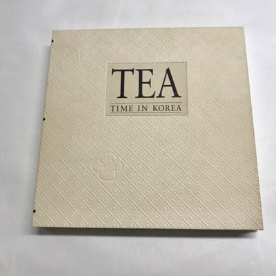
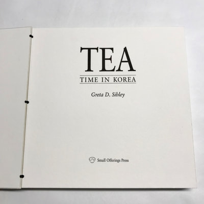
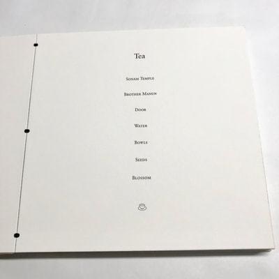
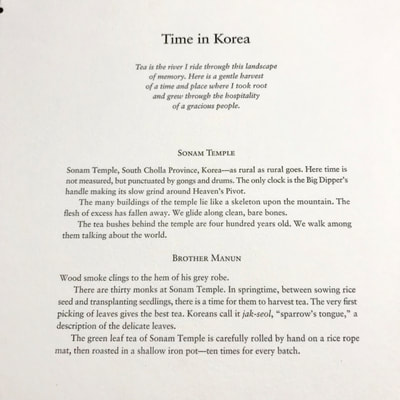
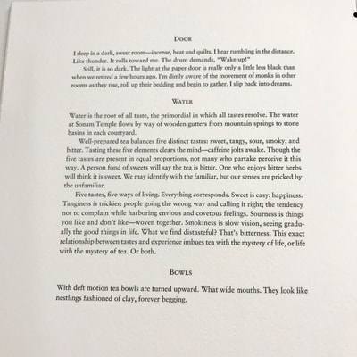
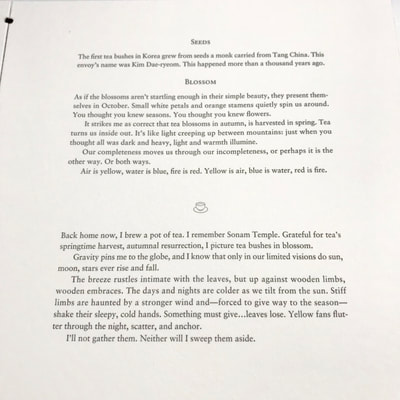
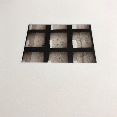
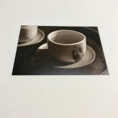
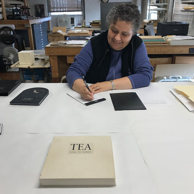
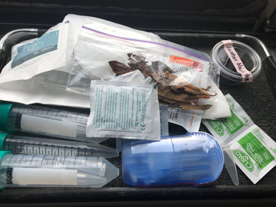
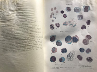
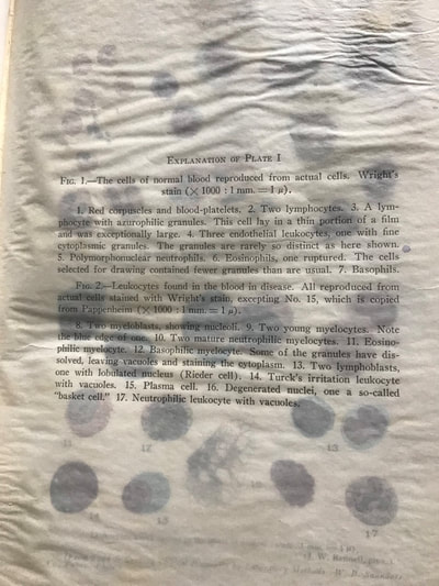
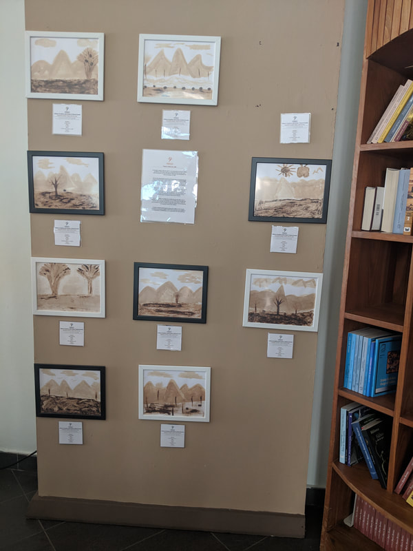
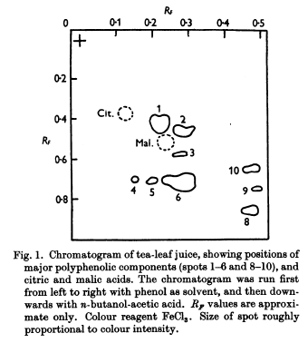
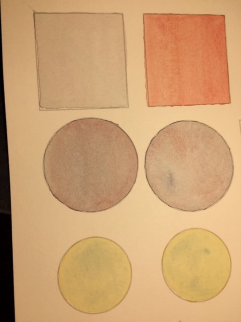
I continue to feel in transition from one studio location to another as I prepare for my 5 week sojourn in Paris, another goal of my sabbatical. I will be there to investigate and photograph Marie Curie’s radioactive journals at the NbF. However, this week I am still in my New York apartment although I did take the train on Sunday to Springfield MA to finish the publication of an artist book collaboration with poet Kim Bridgford (14 years in the making from concept to physical form). While there I came across a book on TEA that our designer, Greta Sibley, had produced in the 1980s after a residency in Korea. It was a moment of chance she had selected this book to show both of us how we might structure a colophon. When I noticed upon closing the book that it was called TEA, it started a discussion on the Bridge collaboration. I have included photographs of that lovely book. In additional to its square minimal format, I was interested in how in the text Greta separated her reflections on that period of time into the separate elements of tea as well as the ceremonies in which she was involved, ie. water, bowls, seeds, blossoms. Her quiet words and evocative photographs presented another aspect of this collaboration with Montse as we dually explore tea from our varying perspectives... the book added a sense of time, as in the tea ceremony one is asked to give up a separate existence and become one with the tea through breath, scent, heat, the cupping of the bowl, the color of the liquid, the quiet, the stillness. It is a vehicle to understand the nature of existence. If the tea, as such, needs to transition from the seeds and ensuing plants to the essence of a carefully prescribed brew, it is this journey seemingly parallel to our own, that is equally as important as understanding the scientific explanation of its molecular composition. My time on Sunday ended with the discovery and purchase of a vintage book on molecular structures and how finding that book coincided with opening the package of lab paraphernalia Montse sent to me. Tools, leaves and brews to explore and experiments to undertake. Montse Last week I had been thinking a lot about tea pigments, their relationship to society, watercolor painting and science. Before I elaborate on my thoughts, I would like to define paper (solid media) and pigment (dissolved in a liquid media). Paper: The paper used in water color is made out of 88-96 % cotton1. In water color, the type or quality of the paper is very important. In more detail, cotton is mostly α-cellulose1, which is a long molecule made out of repeating building blocks (β 1,4 glycopyranose),2 with very little contaminants. Additionally, the paper can have different textures, depending on how it is prepared; hot press is smooth and even, cold press is less smooth, and finally, the rough paper is highly textured. Pigment: According to the Merriam Webster Dictionary, a pigment is: a substance that imparts black or white or a color to other materials especially: a powdered substance that is mixed with a liquid in which it is relatively insoluble and used especially to impart color to coating materials (such as paints) or to inks, plastics, and rubber. The connection between science, society and art lies in the use of paper, pigments and water. In art this is called water color, in science it is called paper chromatography. Below, I will explore three different cases that illustrate the same process but are used for different purposes. The first example came up this week when I was talking to one of my colleagues that came back from a trip to the Dominican Republic. During that trip, she visited the Centro Cultural Perello’ (Figure 1) that had an exhibition called “Papel y tinta de caf” in which the artists (children) created landscapes on paper using coffee as the pigment (Figure2). In science, paper chromatography has been used to separate mixtures into individual components. In fact, in 1950 Roberts and Wood3 used paper chromatography to study the different polyphenols (antioxidants) in tea leaves (Figure 3 shows the result they obtained using paper chromatography). Nowadays, paper chromatography is not used as often. It has been replaced by Thin Layer Chromatography and Liquid Chromatography. Watercolor painting is performed when a pigment (that is either suspended or soluble in water) is transferred into the paper and it is used to create art. Watercolor pigments can be made of inorganic molecules (containing metals) or organic molecules (containing atoms such as carbon, oxygen, hydrogen and nitrogen). The following web page has a comprehensive list of many pigments used in art (The Color of Art Pigment Database). The following figures show how different pigments can diffuse differently on paper. Figure 4 shows various figures that have been painted using watercolors: the first square has been painted using only red pigment; the second square shows a mixture of pigments (blue and red) that traveled evenly, showing a homogeneous smooth color; the third set includes the two middle circles and was painted using a mixture of blue and red pigment (an attempt to make violet), and it can be observed that in the top circle the blue pigment diffused at different rates compared to the red pigment, thus causing a strong blue coloration close on one side and a red coloration on the other side; the final set shows a similar effect, but in this case, the circles were painted using blue and yellow pigments. Jo
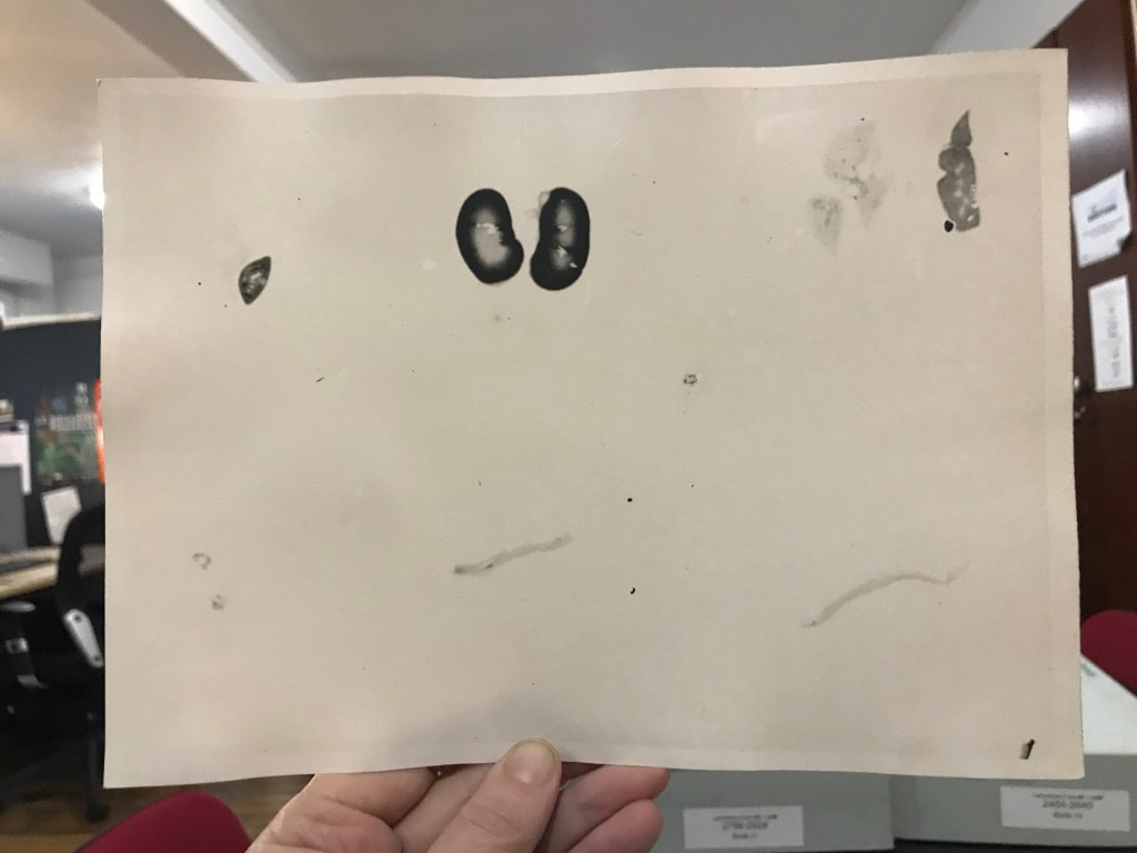
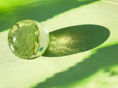
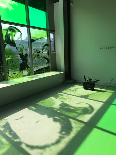
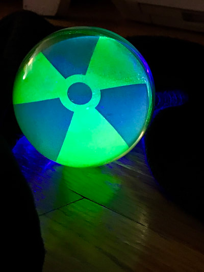
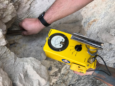
This past week has been a slow one as I am unpacking studio work from recent artist residencies at VCCA and the Anderson Center. In two weeks’ time, I will be in Paris to work on photographing and interacting with the Marie Currie notebooks at the NbF in Paris. During the last seven days, I had a bit of time to reflect on the aspect of stain and molds and my ongoing conversation/experimentation with Montse’s. This seems to be correlating with my unpacking and viewing not only recent residency work but a much more broad-based concurrent unpacking (and divesting) of earlier artwork and exhibition documentation. Stain, aftermath, residue, and imprint keep reappearing in one form or another in all my work. These interests also carry over in to another concurrent project - uranium, its radioactive properties, its implementation, and its aftermath. I am curious to see how these various projects overlap and evolve. What will rise to the top as more expansive themes? I have uploaded some of my concurrent uranium-focused projects to add context to the investigation of the teas. I am hoping before I leave to try some of the innovative approaches to Cyanotype chemistry suggested by Montse - combining ferric cyanide and tea (which as she mentioned might replace the hydrogen peroxide or the ferric ammonium sulfate). An exploration of how iron in blood might react, as in the tea, to the iron in the Cyanotype chemistry. I look forward to the teas and materials Montse is sending to me this week and will forward her my test results when I have a chance to work with them. Also, as a post script... Montse and I have been discussing my coming to Flint to exhibit our work and perhaps engage in experimental sciart workshops with the students. We are currently working out the possible structuring of this interactive work and scheduling logistics. |

