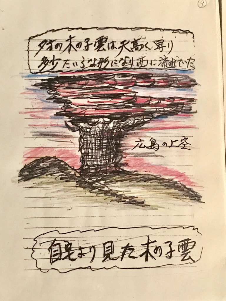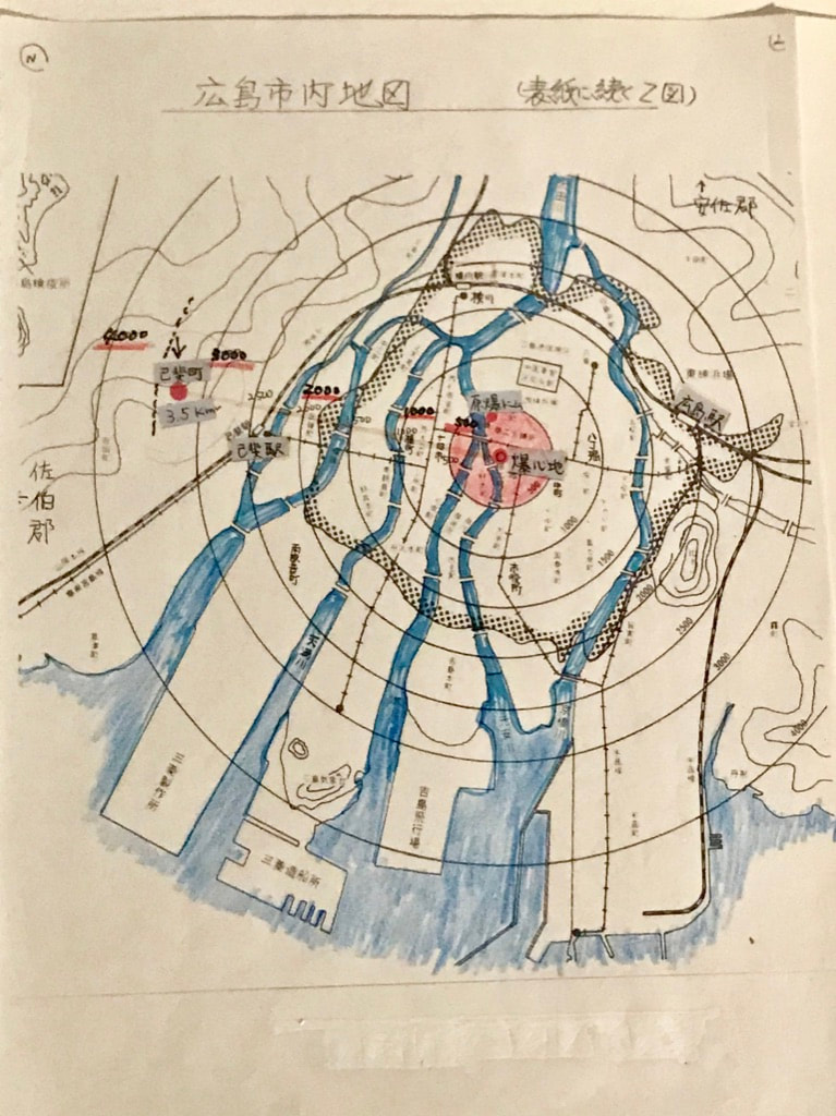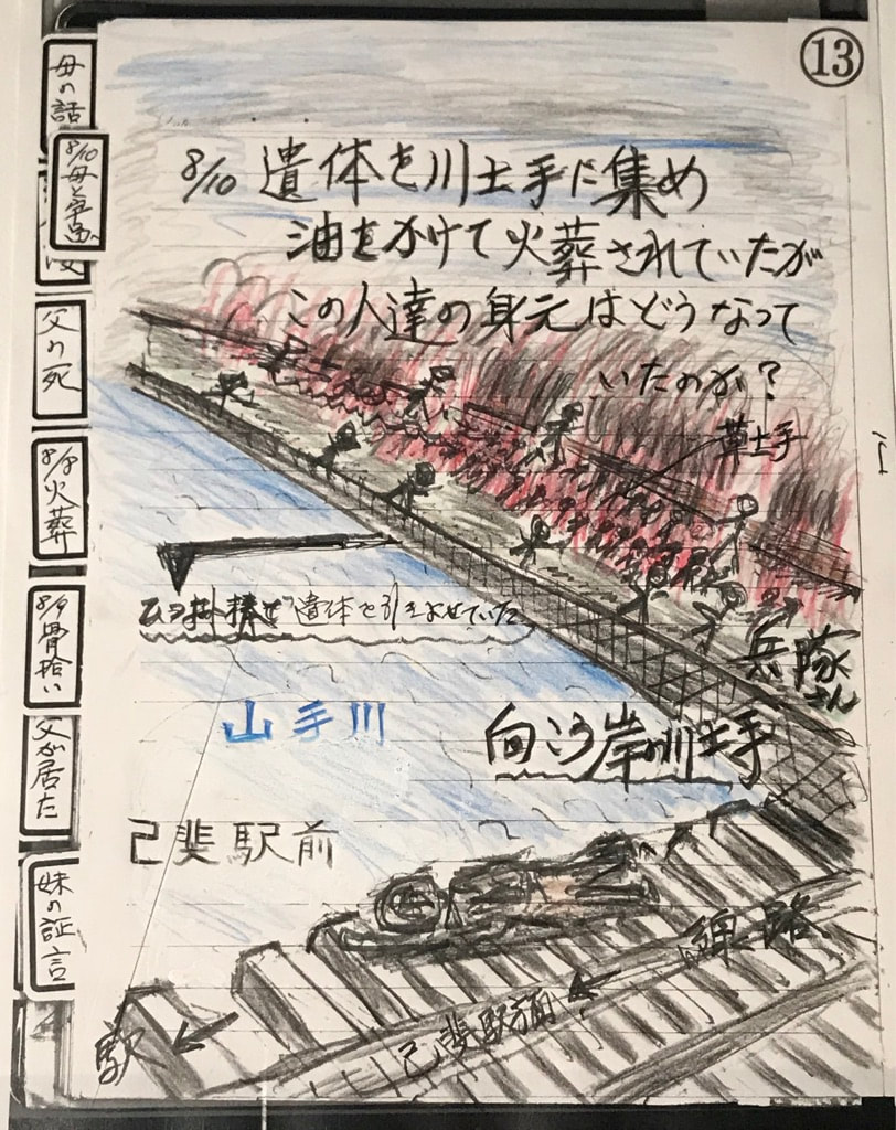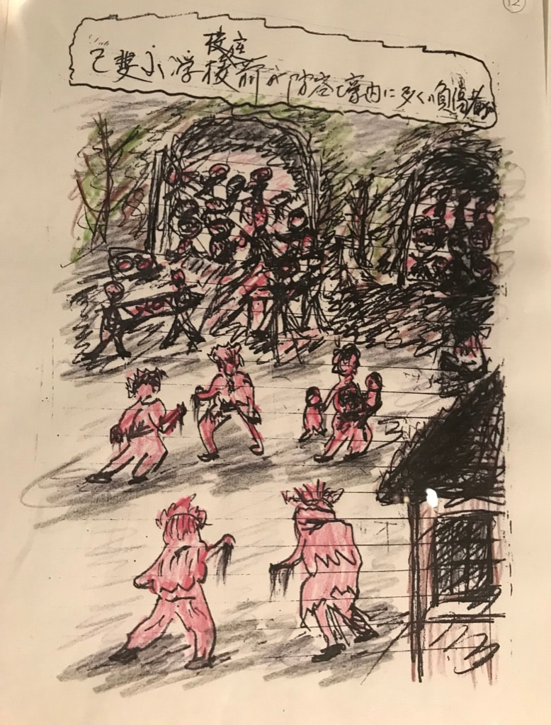
|
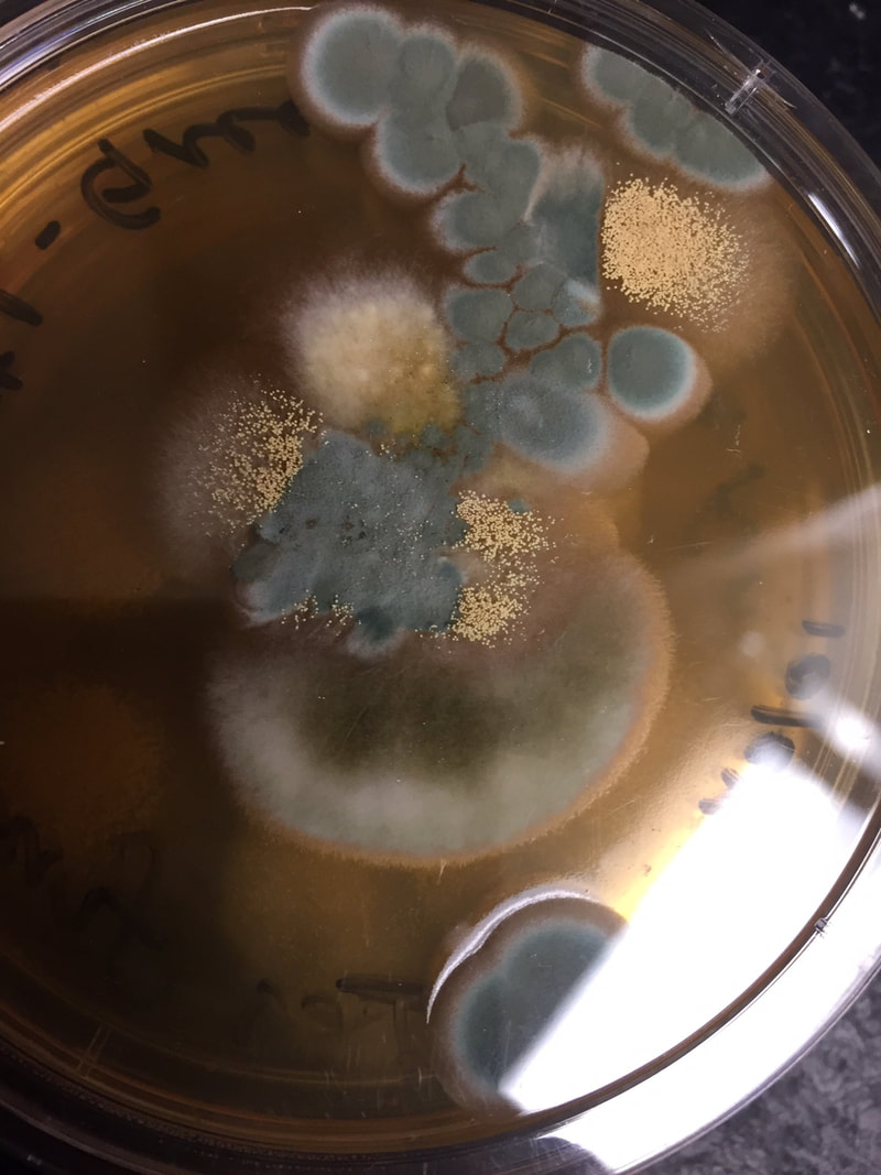
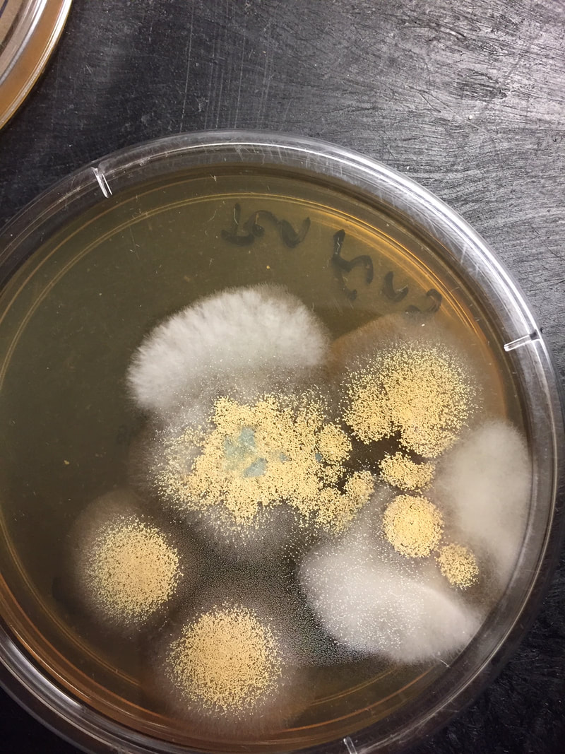
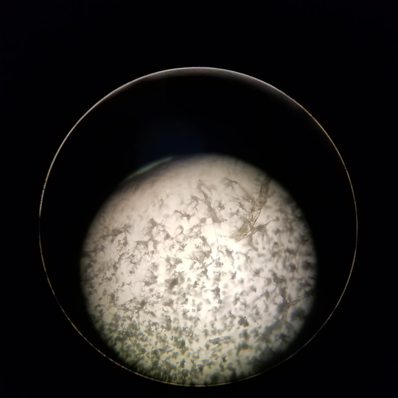
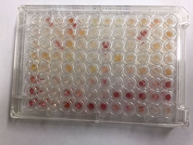
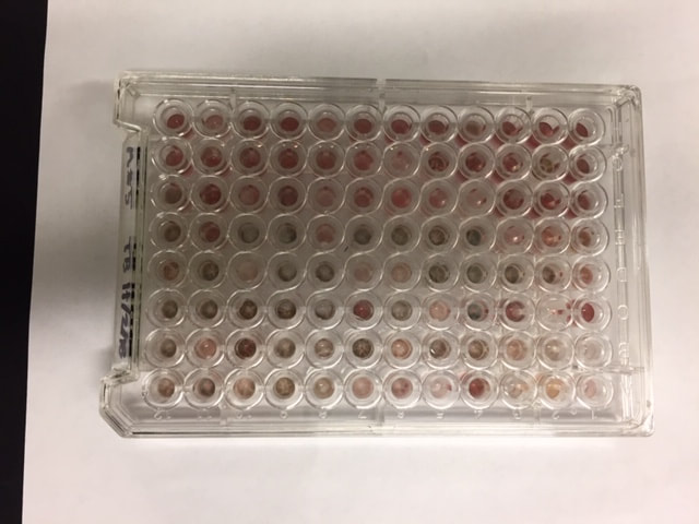
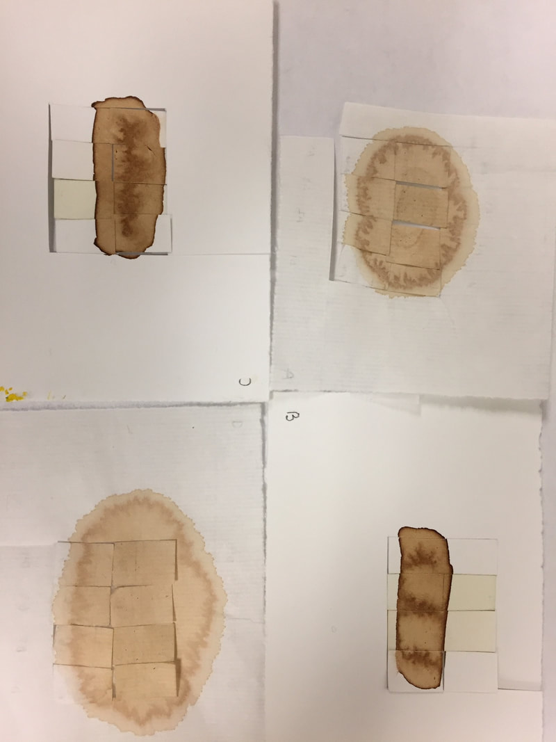
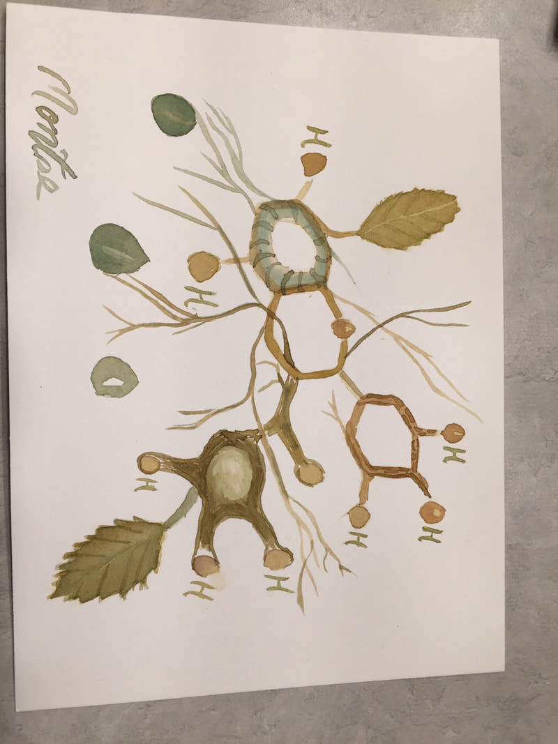
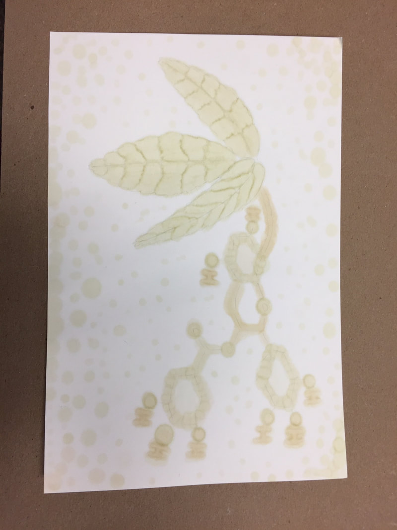
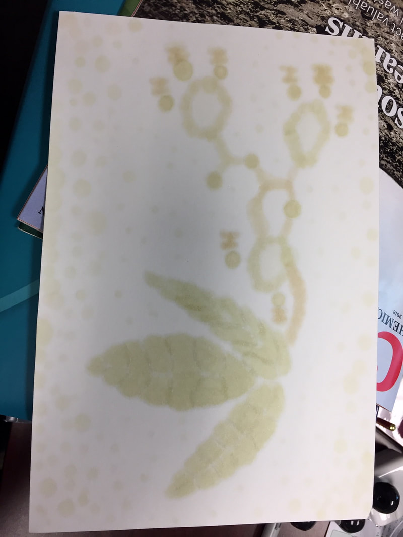
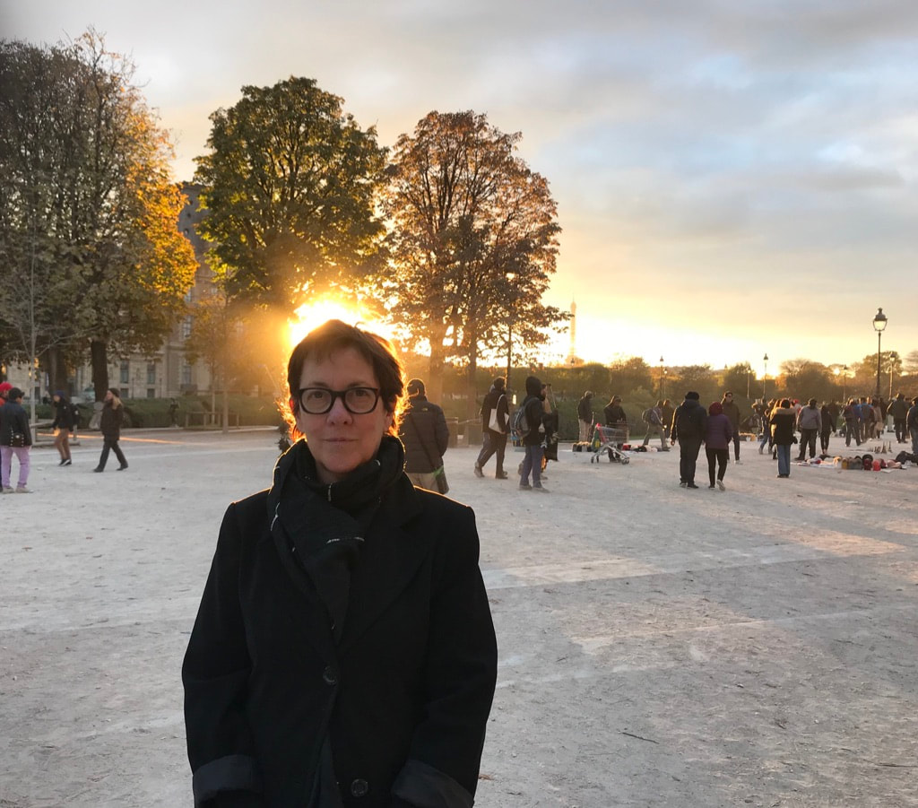
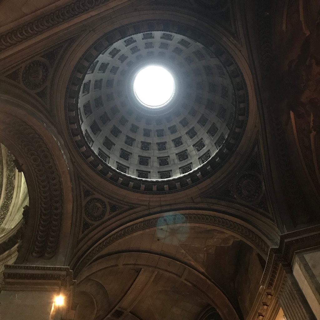
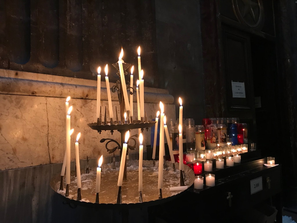
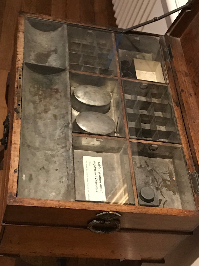
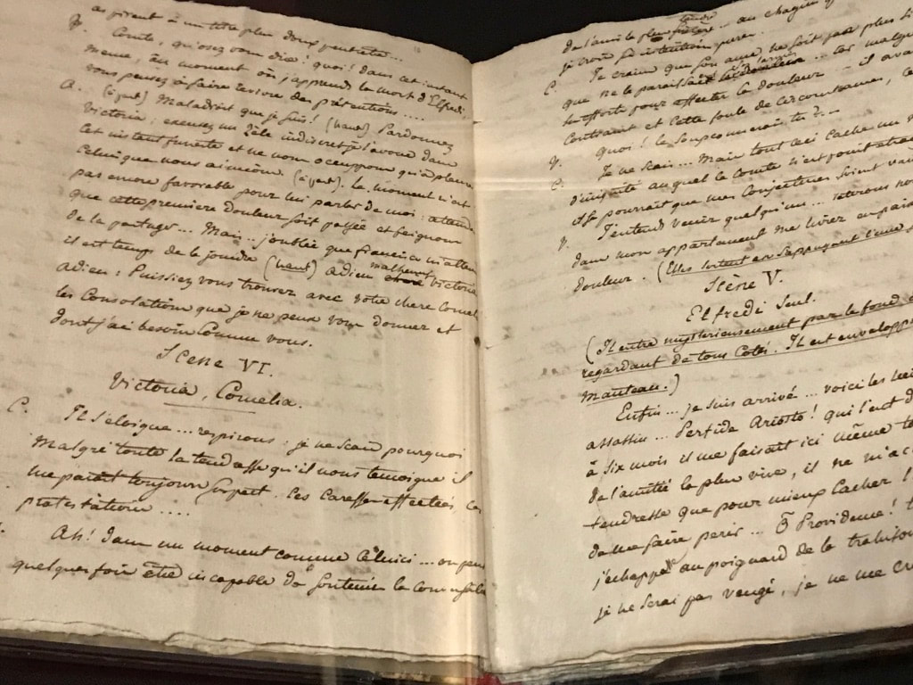
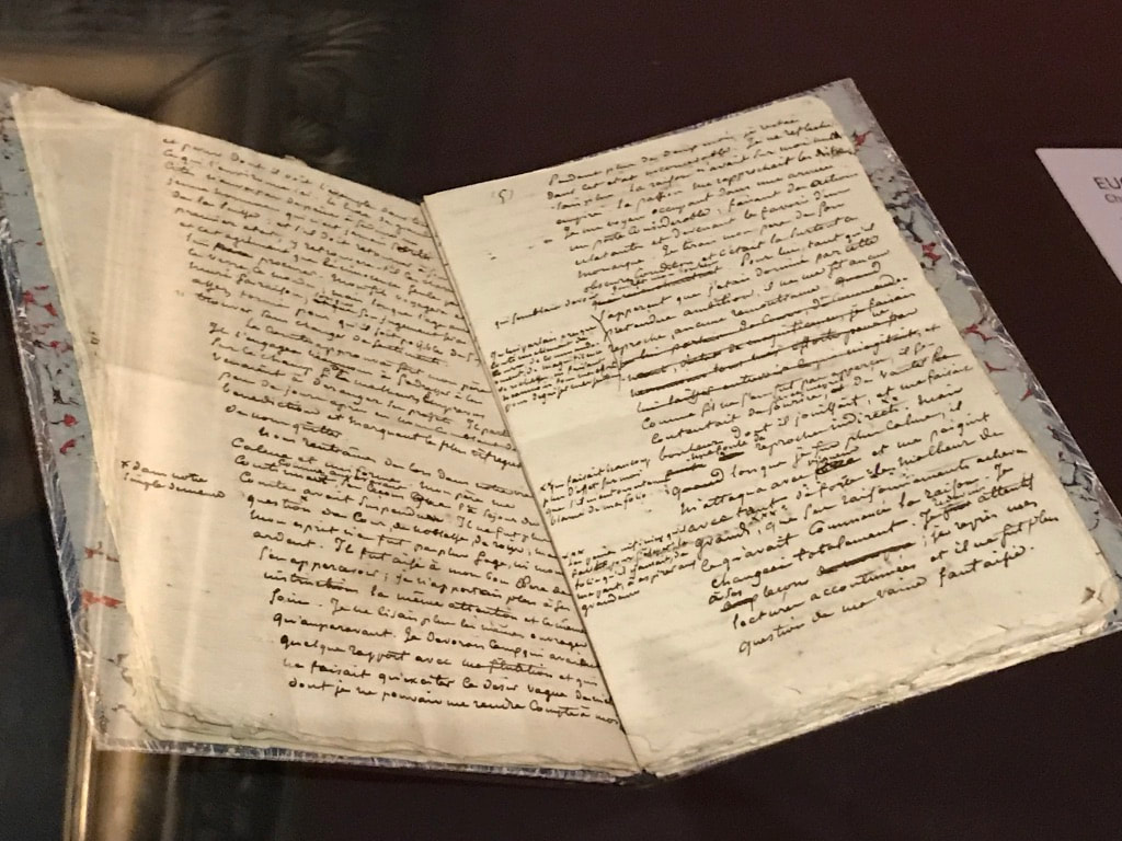
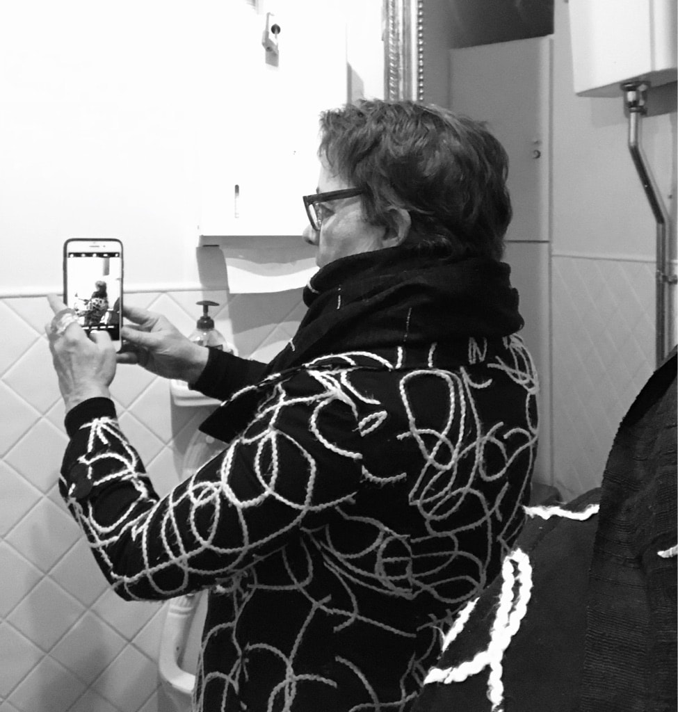
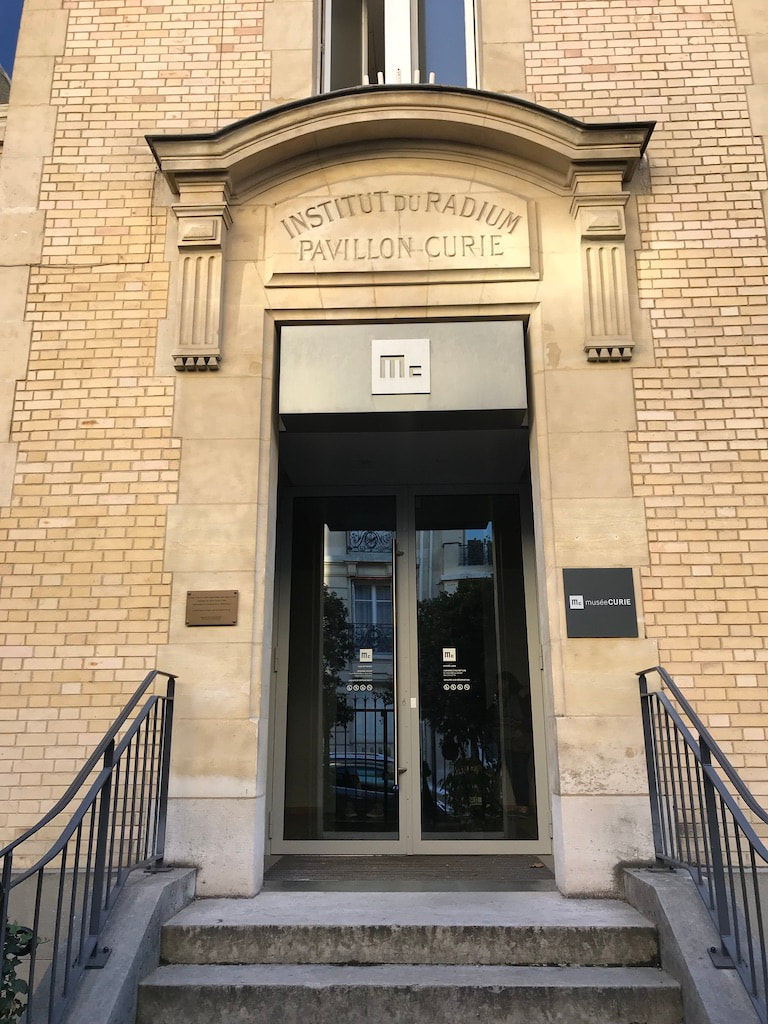
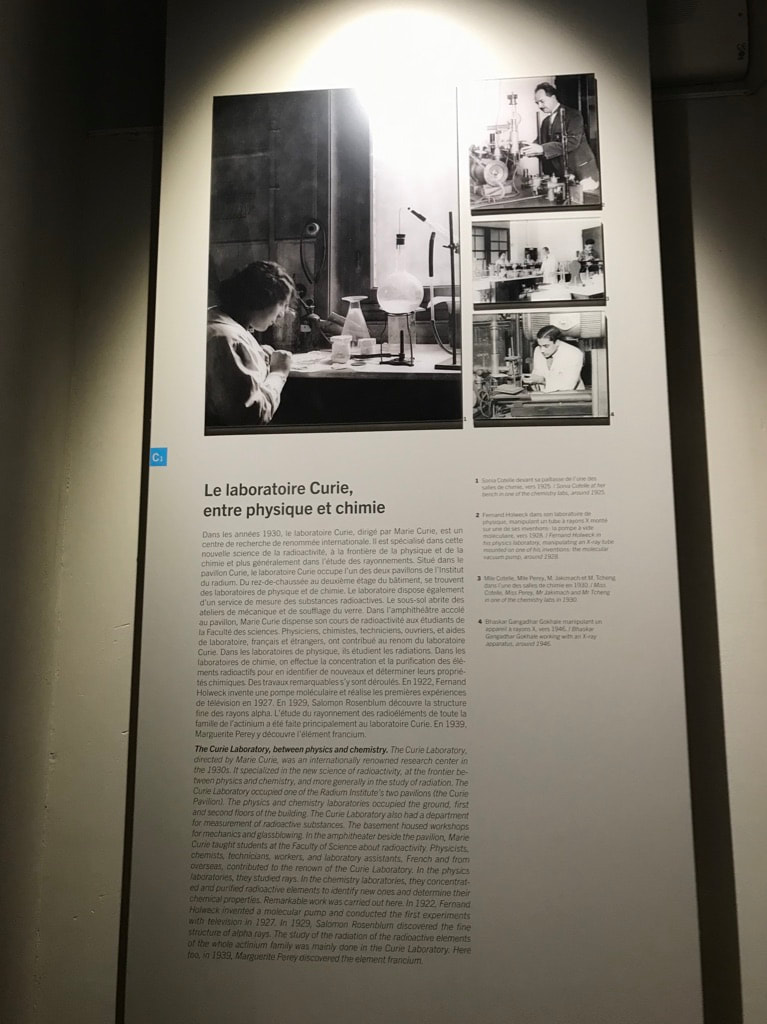
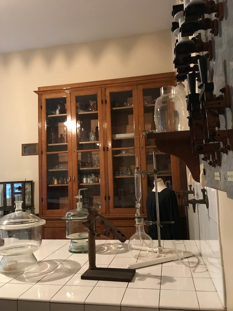
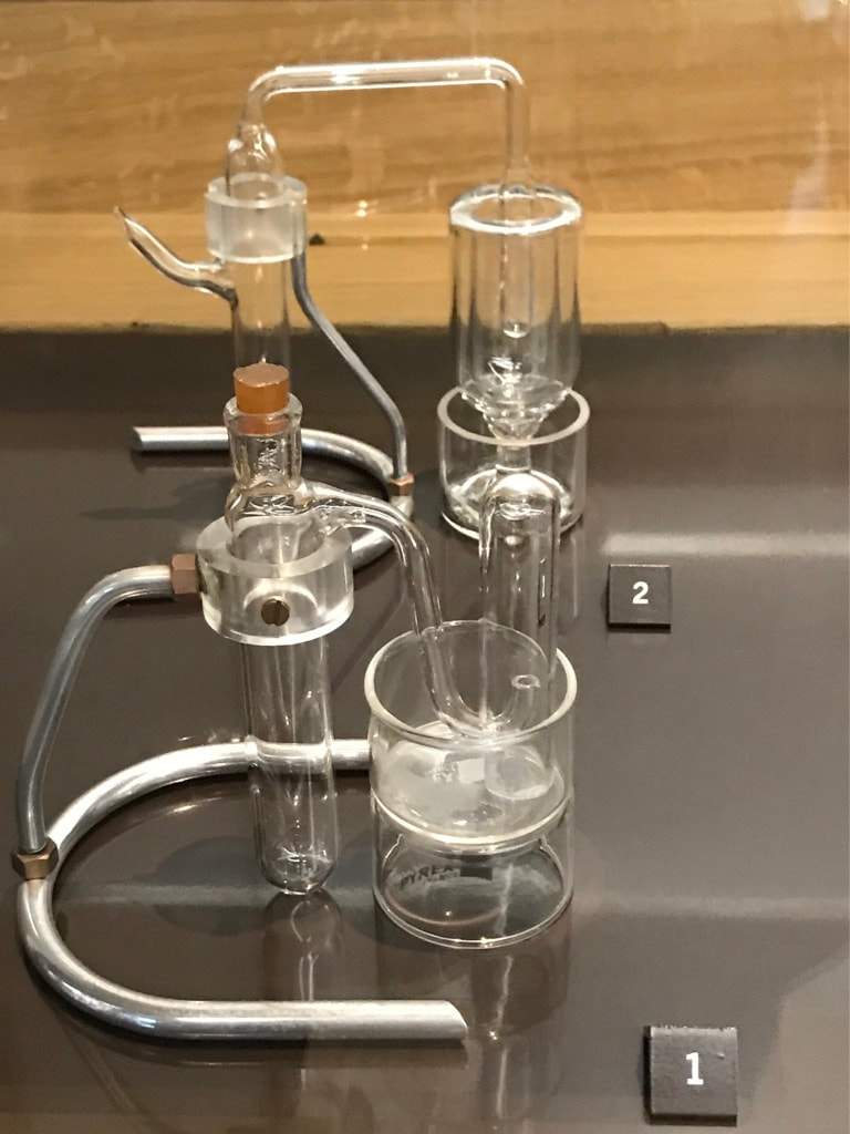
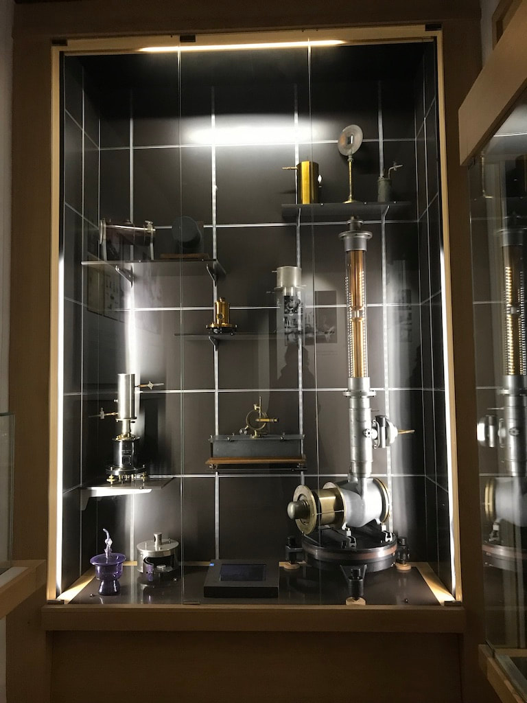
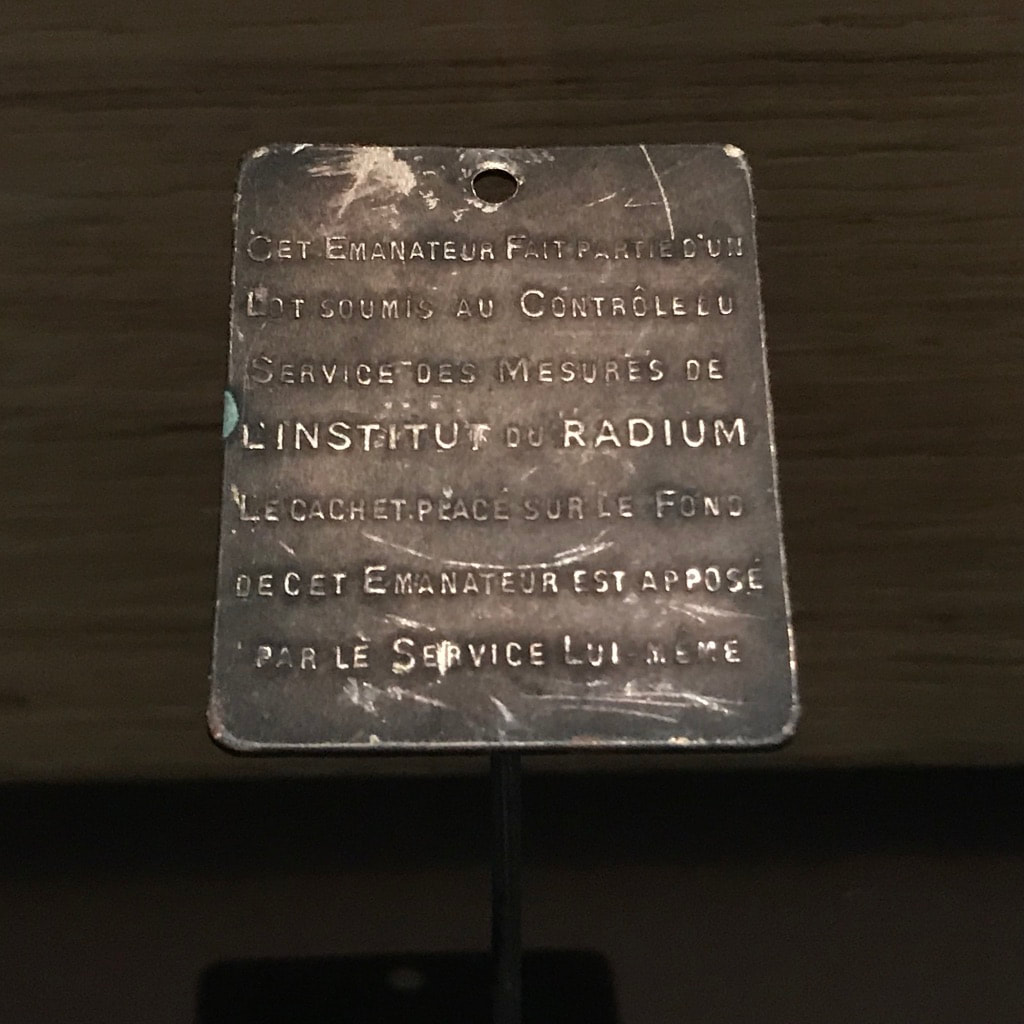
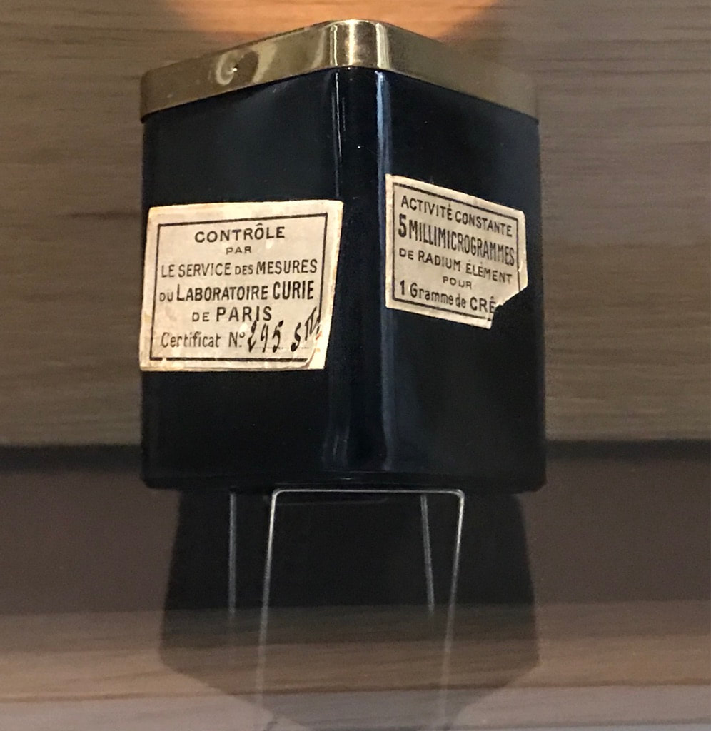
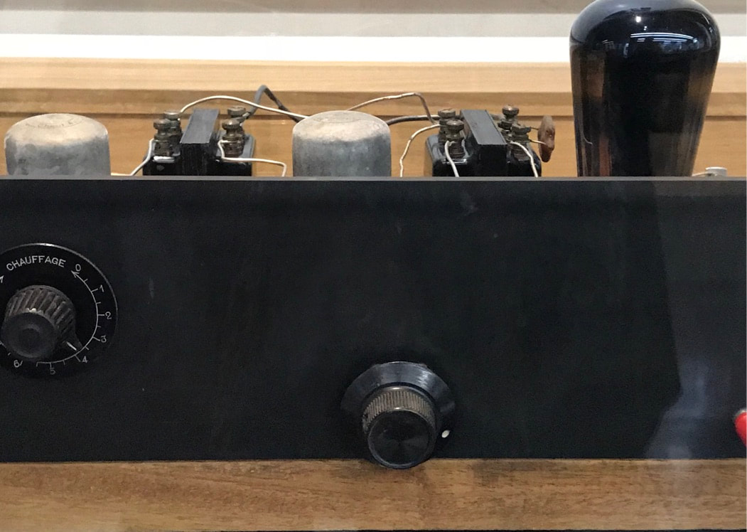
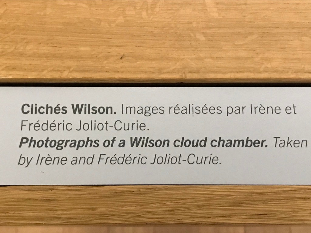
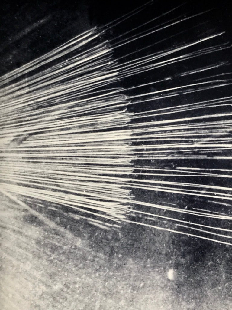
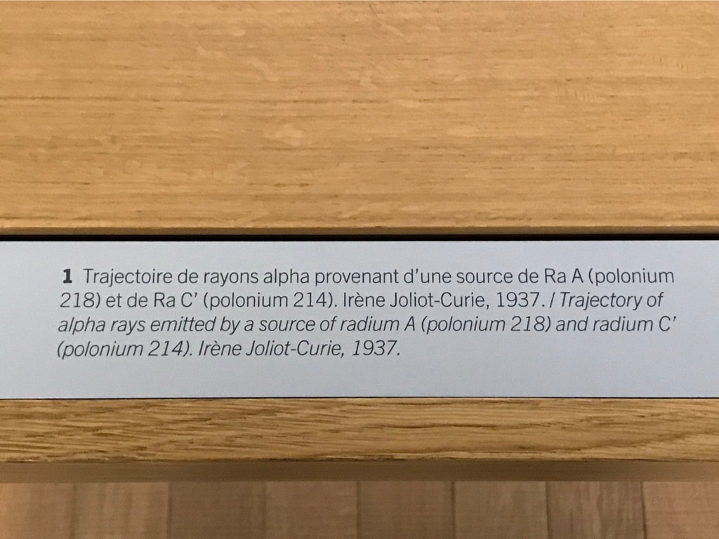
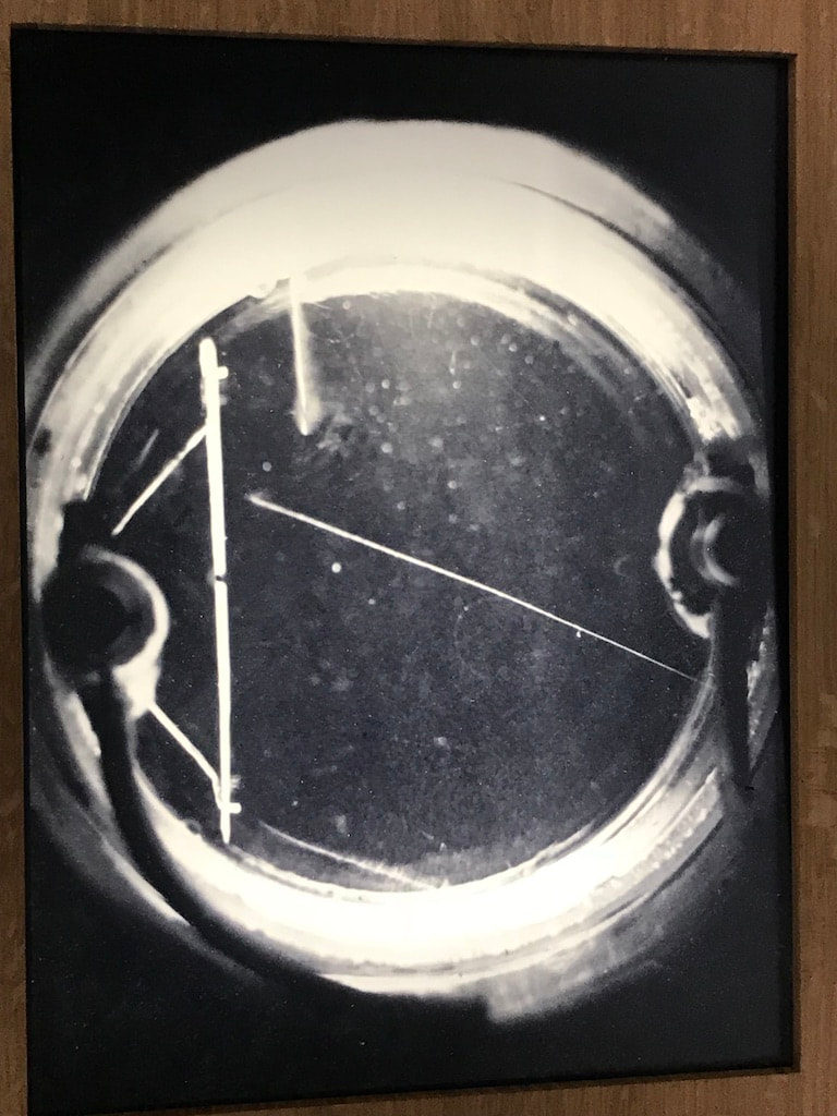
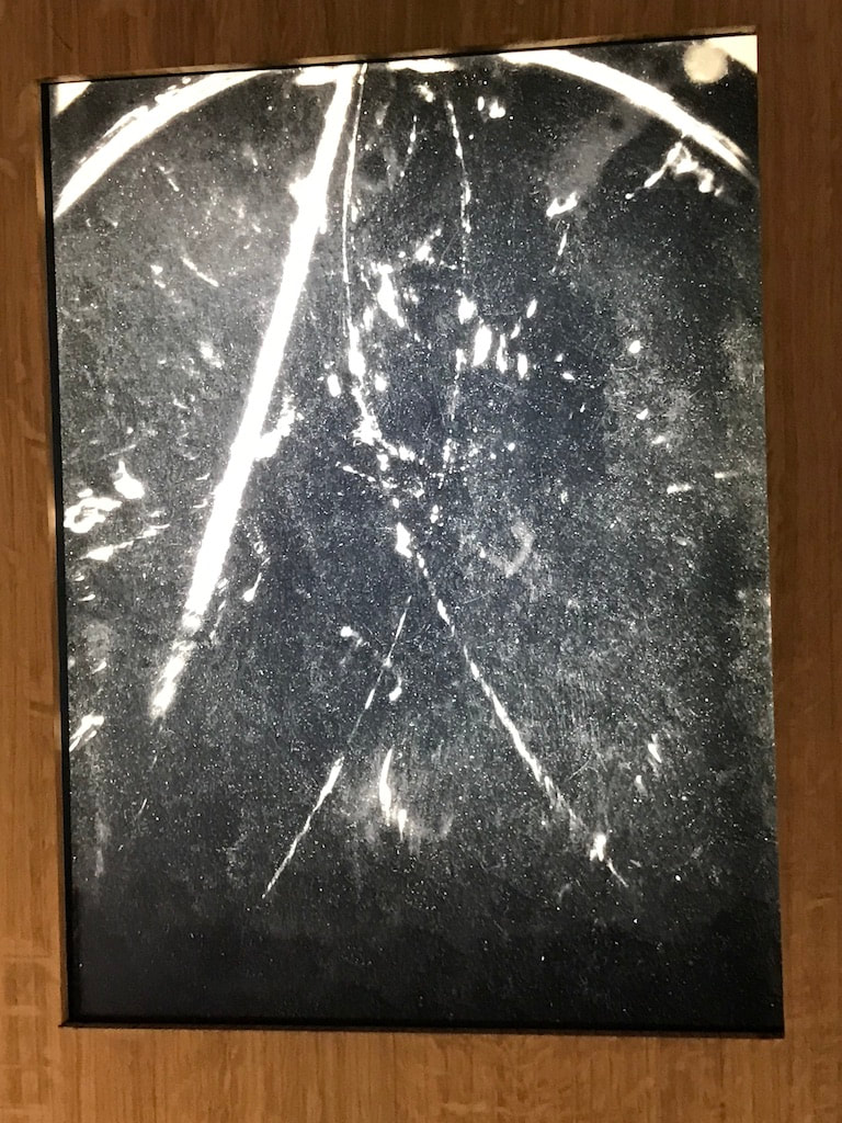
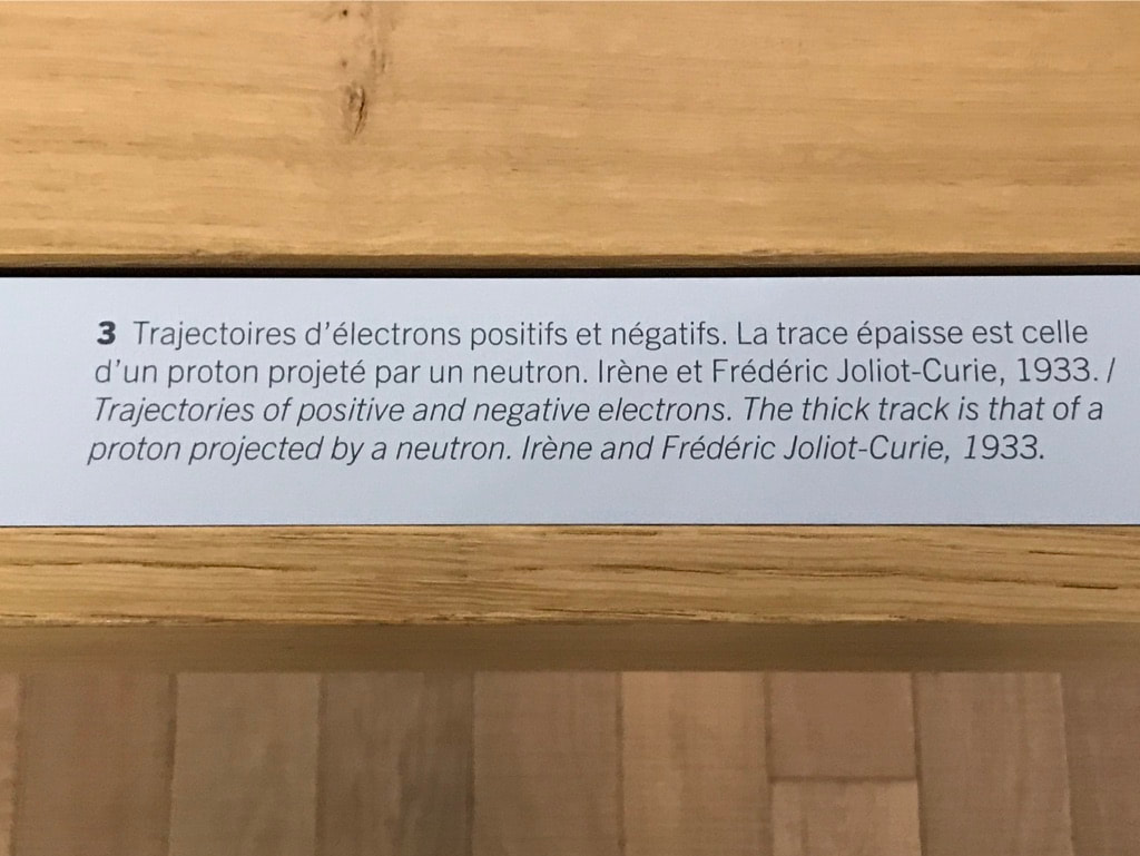
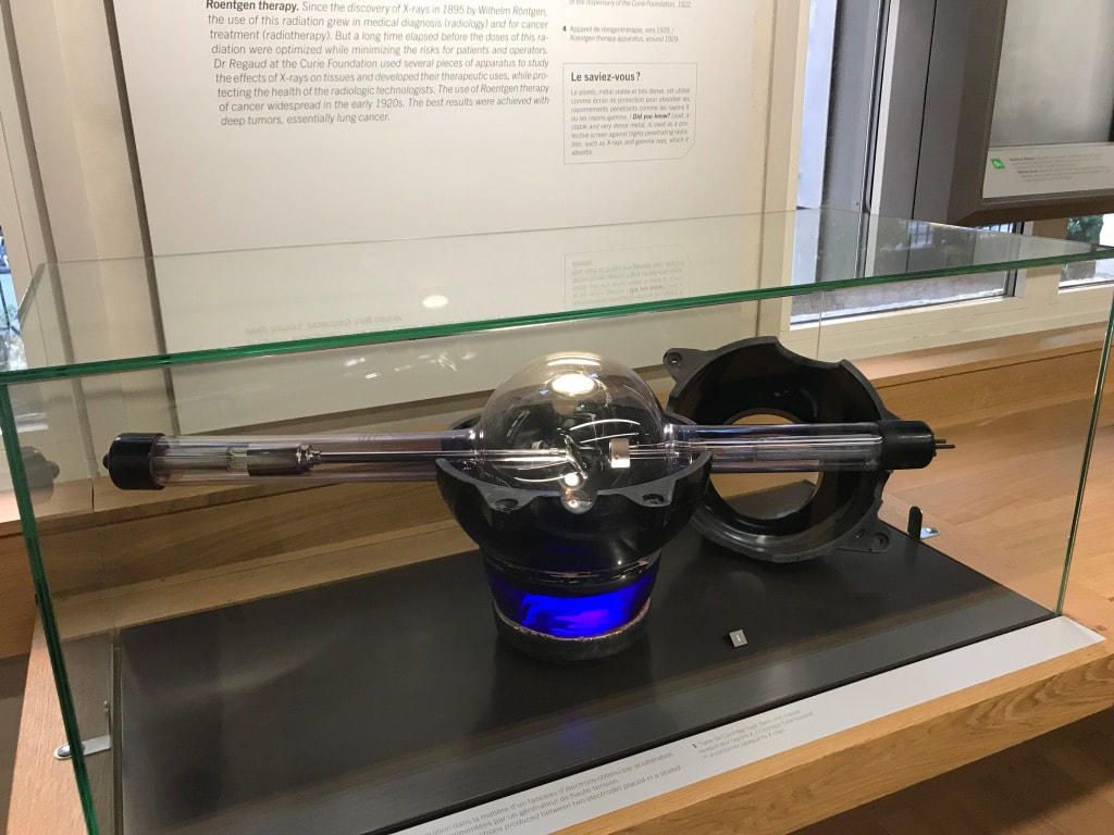
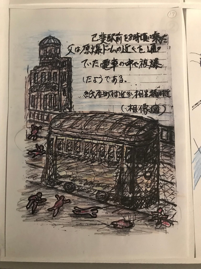
Montse The mold samples are being tested by students in the Microbiology class. First, the mold was plated (to separate individual types of mold) (Figures 1-3). Pictures of the molds under the microscope are shown in figures 4-6. To identify the molds present in those samples, the FF MicroPlateTM from BIOLOG was used. The FF MicroPlateTM test panel provides a standardized micromethod using 95 biochemical tests to identify / characterize a broad range of fungi including both filamentous and yeast forms.1 Liquid cultures containing individual colonies were grown, and were used in the FF MicroPlateTM, figures 7-9 show the pictures of the 96 well plates. Analysis of all of the data collected to determine the type of fungi is still underway. The experiments performed on the tea stain samples are mostly complete. Each one of the stains was divided into eight sections that were equal in length. Each one of those sections was tested under unique conditions. The reassembled tea samples are shown in figure 10. In conclusion, using the FTIR information, both papers are made of cellulose (see previous blog for details). However, they differ significantly in regard to the thickness and density of the cellulose fibers as well as the surface tension between both papers (see previous blog for details). Another very interesting difference is the apparent variation in hydrophobicity; the thick paper appeared to be a lot more hydrophobic compared to the thin paper. This was an unexpected observation; the reasons for this are unclear and under investigation. Also, Jo and I have continued to think of ideas and activities that can be integrated into an exhibition. One of our ideas is to explore and expand on the use of tea to create something. This idea has been previously explored by other artists; a few examples are shown in the links below (Link 1-5). In order to incorporate the science aspect, I decided to explore a similar idea to Gerard Tonti (link 1), but use the previous extracts (see previous blog for details) to create a watercolor paint. To compare how the tea paint would look versus the extract paint, I composed two drafts: one using real watercolors (figure 11) and one using extracts (figure 12 front of paper figure 13 back of paper). In both, I attempted to draw the tea leaves and one of the most important antioxidants in tea: epigallocatechin gallate. Links: Link 1 https://www.dailymail.co.uk/news/article-2452341/Putting-TEA-art-Artist-swaps-paint-hot-drinks-create-intricate-portraits.html Link 2 https://www.huffingtonpost.com/2015/02/05/tea-bag-portrait-art-red-hong-yi_n_6622180.html Link 3 http://redhongyi.com/portfolio/untitled-tiger-with-tea-leaves/ Link 4 https://mymodernmet.com/miniature-paintings-tea-bags-ruby-silvious/ Link 5 https://www.brandsouthafrica.com/play-your-part-category/play-your-part-news/women-recycle-tea-bags-to-make-art 1. www.biolog.com, FF MicroPlateTM Manual. Jo As mentioned in my last entry, this week I was visually researching/getting inspiration from visiting in Paris with other artists and curators a number of art and science museums as well as searching out obscure cultural locations and events. I have a number of examples of locations and works which have inspired me and have labeled and arranged them to create a kind of spontaneous and chance-based narrative (a type of artform, perhaps). Also following is a copy of my email communication with the Curie Museum staff/curators as I first visit and then try to gain entry to the collections, to photograph with a stereoscopic camera the Curie lab equipment and notebook pages: Dear Professor Yarrington, First of all, thank you for your interest in our collections ! Before giving you an answer I need to discuss your request with the head of the historical resources department, who will only be available next week. I will contact you as soon as I have more information, sorry for the delay. Best regards, Aurélie LEMOINE Aurélie LEMOINE Archiviste Musée Curie - UMS 6425 CNRS/Institut Curie adresse postale : 11 rue Pierre et Marie Curie 75248 Paris Cedex 05 adresse de consultation archives : 21 rue Tournefort 75005 Paris Tél. :+33 (0)1.56.24.55.49 [email protected] Rejoignez-nous sur Facebook De : Yarrington, Kathryn J. <[email protected]> Envoyé : lundi 5 novembre 2018 15:11:38 À : Klapisz Adrien Cc : [email protected]; documentation.musee Objet : Marie Curie archives Dear Adrien, It was wonderful meeting you last week and I thank you for your kindness in spending so much time at the end of the day to help me with my visual art research and process focused on Marie and Pierre Curie’s discoveries. As mentioned to you when at the Museum, I am in Paris for 5 weeks from late October to December 2018 to research and photograph at the National Library and at your museum, specifically focusing on Madame Curie and her journals.. By way of an introduction to your other colleagues at the Museum, my name is Jo Yarrington and I am a visual artist and full professor at Fairfield University with a primary focus in printmaking and photography, although I also have been developing work in comprehensive installations which have included glass production and artist book series. My website is www.joyarrington.com>. I live in New York City and have been awarded a yearlong sabbatical leave, 2018-2019, to continue my focus on uranium as product, process and political “code”. I have two solo exhibitions coming up in spring 2019 in museums in New Jersey and China, both using uranium as product and metaphoric lens. Because my work is multi-disciplinary, this focus has taken me from documenting Nuclear Power Plants in the United State and Europe to more recently photographing with a small group of collaborators abandoned uranium mine sites in the state of Utah and the Navajo Nation (located in southern Utah) where we have been researching the current politics and local history (including physical artifacts found at the mines). Our photographic work also extends into alternative photographic processes and we have been using uranium to make uranium prints (a process my colleague, photographer Morgan Post, recently resurrected from a mid-1800’s formula). Using this formula, we employ either negatives from images taken at the mines or, specifically in my case, as photograms derived from objects, a la Man Ray. While in Utah, among other mines, I visited the Temple Mountain Mining Complex from which uranium was extracted and shipped in the late 1800’s to France to be used by Madame Curie for her experiments with pitch blend extraction. As mentioned, I would be interested in the following help. My aim is to photograph laboratory objects and manuscript pages not yet digitalized. For this, I am using a vintage stereoscopic film camera (commonly known as a Viewmaster) to photograph the Curie notebook pages and laboratory objects, thus creating an optical dimensionality. I will have an example of one of the Viewmaster cards and a Viewmaster to examine with me, to aid in clarification. Famed photographer Steven Shore also used this method in some of the groundbreaking series he did in the 1970’s. Steroscopic photography relates to the historic time in which the notebooks were written by Marie Curie and provides a metaphoric resonance, a way to look at scientific discoveries, as Marcel Proust would say, through “two lenses”. Although digitalized photographic reproductions probably are available of most items, it would compromise the integrity of that central idea for me to not be able to photograph the existing pages and/or objects, as outlined in the following. You mentioned that I should be specific since different curators are responsible for each area in which I wish to view and photograph: * Notebook pages: To view and subsequently photograph non-digitalized Curie notebook pages currently in the archives * Scientific images: To view and subsequently photograph the science-based images such as the photogaphs of the Wilson cloud chamber, the trajectory of alpha rays emitted by a source of radium, trajectories of positive and negative electrons and any other derived image in which special equipment was used to explore reactions and emissions. * Glass-based laboratory equipment: To view and subsequently photograph glass laboratory equipment, especially that which was used to extract radium or was used as a health-based light emission (such as the black light tubes). I would be most grateful for any help you can offer to move forward with my research. I am available this coming Wednesday and Thursday (8, 9, 10 November) to visit the Museum again and perhaps discuss with each curator what I might be able to see and what I might, at a later date, be able to photograph. I look forward to hearing from you at your earliest convenience. Sincerely yours, Jo Yarrington Professor of Visual Art Fairfield University United States From: Klapisz Adrien <[email protected]> Date: Friday, November 2, 2018 at 4:48 PM To: Kathryn Yarrington <[email protected]> Cc: "[email protected]" <[email protected]>, "documentation.musee" <[email protected]> Subject: [External] Marie Curie archives Musée Curie : http://www.calames.abes.fr/pub/curie.aspx#details?id=FileId-480 http://www.calames.abes.fr/pub/curie.aspx#details?id=FileId-450 National Librairy : Bnf https://archivesetmanuscrits.bnf.fr/ark:/12148/cc7376q https://gallica.bnf.fr/services/engine/search/sru?operation=searchRetrieve&version=1.2&query=%28dc.title%20all%20%22Pierre%20et%20Marie%20Curie%22%29&keywords=Pierre%20et%20Marie%20Curie&suggest=1 archives et informations : "documentation.musee" <[email protected]>
0 Comments
Leave a Reply. |
Montserrat Rabago Smith
Jo Yarrington |