SciArt Initiative

  • Mission
  • Program Archive
    • Events
    • Exhibits
    • Residency
    • Colloquium
    • Magazine
  • Mission
  • Program Archive
    • Events
    • Exhibits
    • Residency
    • Colloquium
    • Magazine

Dana & Richelle: Post-residency testimonials

2/25/2016

0 Comments

 
0 Comments

Dana & Richelle: Final week!

2/2/2016

0 Comments

 
Dana's update:

Hello, hello!  This week for The Bridge, I sent Richelle three more short texts to illustrate.  Instead of focusing exclusively on my research in these new texts, I decided to take a “myth busters” approach.  I’m debunking three neuro-myths.  I am very much looking forward to see how Richelle decides to put these texts into images!  Here’s a short version of some of the neuro-myth text:
 
Myth 1: Analytical people are left-brained and creative people are right-brained.
 
The entire idea of being left- or right-brained has no basis in science.  Multiple studies have demonstrated that analytical/creative people don’t have any sort of enhancement of connectivity or activity in one select hemisphere of their brain.  Anatomically speaking, the brain does have two hemispheres that are connected by a small area in the middle called the corpus callosum.  However, these two hemispheres are normally equally active in healthy people.
 
Myth 2: I can’t learn anything else because my brain is full.
 
A brain is a plastic, dynamic structure that continues to remodel itself throughout your whole life based on experiences.  Scientists sometimes refer to this as experience-dependent learning.  When you learn or have new experiences, your neurons can change how they connect with each other by strengthening or weakening synapses.  Synapses are synapses across which one neuron sends a message to another.  In general, the more two neurons communicate, the stronger their connection will be.  Your brain strengthens connections it learns are important based on experience, and gets rid of connections it learns are unneeded.  When you’re born, you have a ton of extra connections in your brain because your brain is new and you haven’t refined it yet.  As you get older, experience life, and learn to do things, your brain starts to prune away the synapses it doesn’t need, and strengthen the important ones.  Fun fact: Current research estimates that there are somewhere around 100 trillion synapses in the human brain. 
 
Myth 3: Humans only use 10% of their brains.
 
No! This is so false!  It’s safe to say that your whole brain – or most of it – is working for you pretty much all the time.  Different regions of the brain control our various abilities that keep us alive and functioning.  For example, your brain stem controls basic, critical functions like your breathing rate, temperature, heart rate, etc.  In other words, if you are alive, then your brainstem is working.  Other parts of the brain are responsible for things like thinking, planning, seeing, thinking about what we see, helping us walk, keeping us upright, letting us interpret our environment, and formulating appropriate behavioral responses to what’s going on around us.  Whether or not you actively “try” to think about something, your brain is getting it done.
 
In addition to sending neuro-myth busting text to Richelle, I printed some new Purkinje cells and dendrites to bring to The Bridge Symposium on February 17th.  I’m amazed and excited by all the interest I’ve been getting in these prints! 
 
​Here are some of my digital prints: 
Picture
Picture
Picture
Picture
Richelle’s Update: 

Two more weeks until the Bridge Symposium! Right now I am creating an illustration to her text “What are other examples of the Perkinje pattern in nature?” This is one of my favorite questions to ponder because the branching patterns within the Perkinji cell take place in many social, biological, and technological systems. Dana and I discussed other networks that contain similar structural properties. Some examples include: deltas and streams, Facebook diagrams, tree branches, coral, antlers, lightening, family tree charts, and more. 

Prior to the Bridge Residency, seeking patterns that connect all life inspired a 365-day project called Networked Life. I believe that by taking a closer look at patterns in nature, we can more closely understand how this design impact our lives as evidenced in networked social relationships, organizational structures, and communication methods. 
Picture
Networked Life, 365-day project illustrating a network per day completed 2013. Here are 15 selected networks that have similar structural properties to the Perkinje cell.
​Below are images of new materials that will be incorporated into a single drawing. I am compiling and layering this imagery together to reveal common formations within disparate topics. I look forward to exhibiting a series of 10 complete drawings at the Bridge Symposium at the School of Visual Arts.  These drawings will also be included in a series of Science-Art pop-up exhibitions throughout Seattle with support from 4Culture.  
Picture
Brainstorming content that looks similar to the architecture of a Perkinje cell.
Picture
Other forms that mimic the Perkinje cell formation.
0 Comments

Dana & Richelle: Week 17

1/26/2016

0 Comments

 
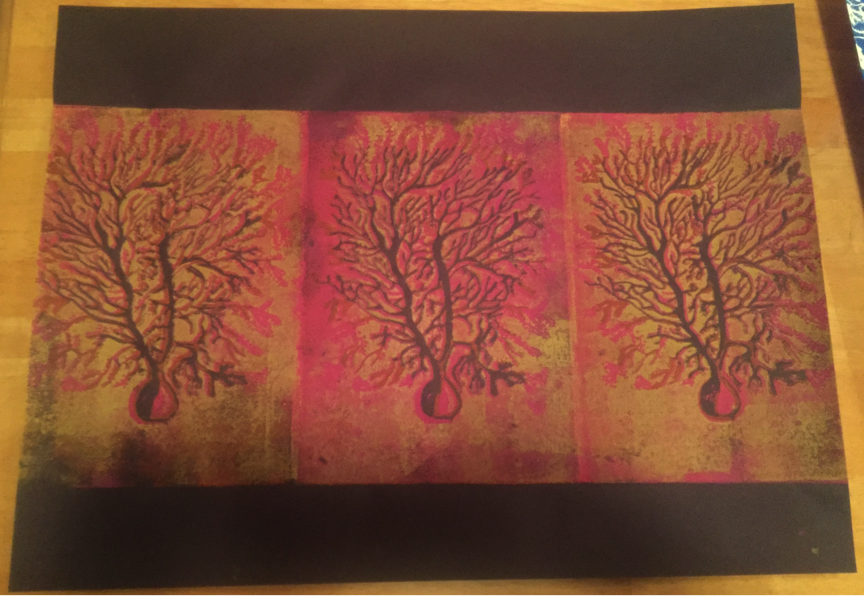
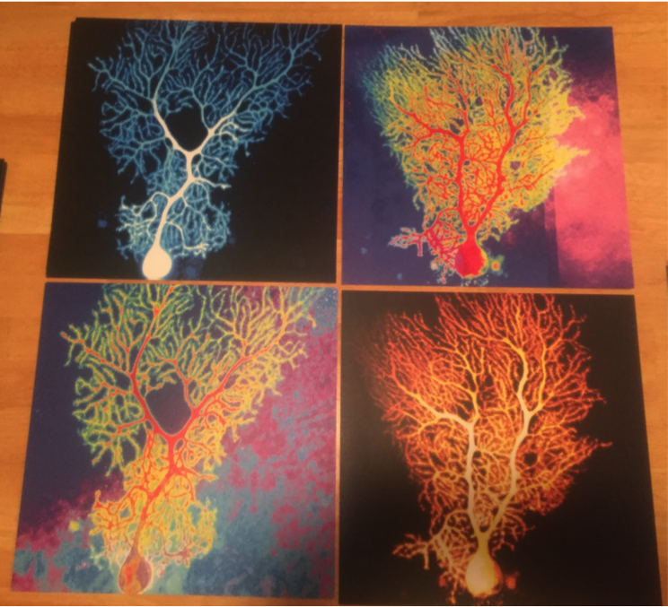
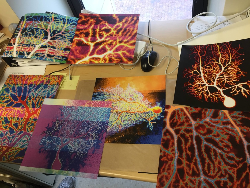
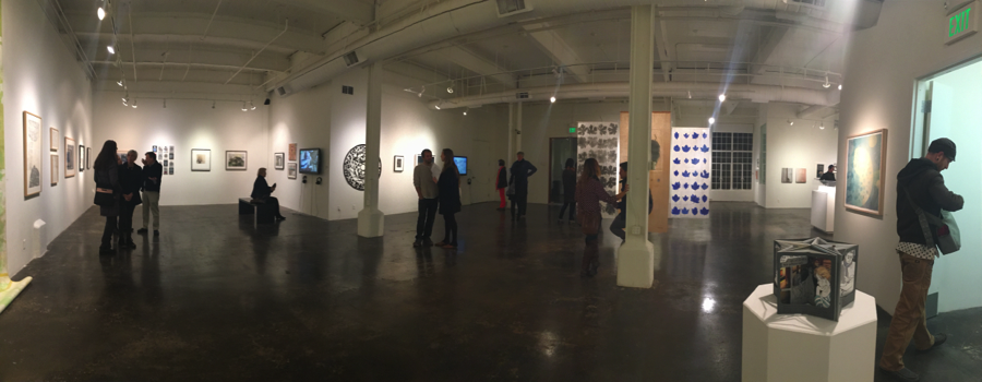
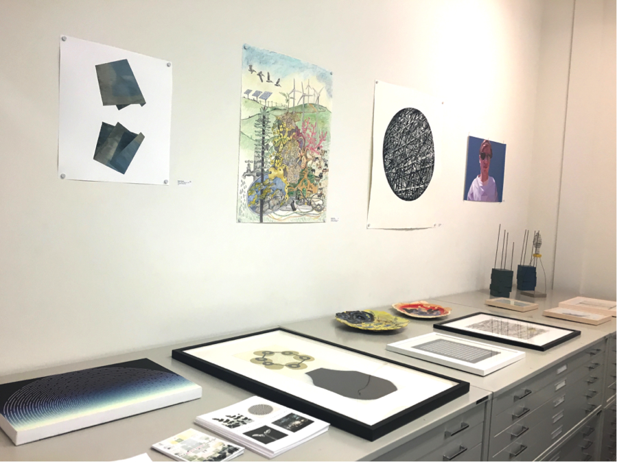
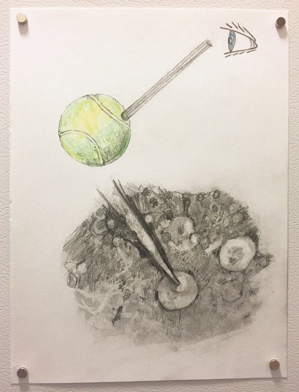
Dana's update:

This week, I’m trying to wrap everything up for The Bridge to get ready for the symposium in New York.  To get ready, I’ve been working on three main things: talking with Richelle about hanging art, and editions and pricing, and finalizing the text for our collaborative project.
 
This week, when Richelle and I talked via Google+, she explained how to hang lightweight art on walls without damaging the art.  She has this cool method of putting nails into walls and then placing magnets over the head of the nail.  The art hangs suspended between the nail and the magnet.  I’m pretty sure my prints will be light enough to try this.
Picture
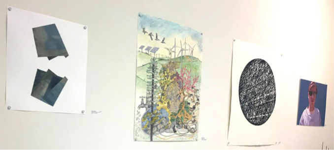
Here are some examples of Richelle’s artworks with the nail/magnet hanging technique.
My first set of prints from last week’s post was a test set.  Now that I have printed these, I can select more neurons and dendrites to print, and maybe even print multiples of some of these images.  Richelle explained how editions work in printing, and recommended that I make about 20 or 25 prints of each.  Richelle explained to me that the funny thing about digital art is that it’s hard to define what is an original vs. a copy.  Since the exact same thing can be printed multiple times, she told me digital artists use editions to designate a certain number of original prints. 
She showed me how to sign the back with the edition number.  While these may all seem like commonplace logistical details to a full-time artist, this information was all new to me, so I’m really glad to have Richelle and Julia to ask about these things. 
Picture
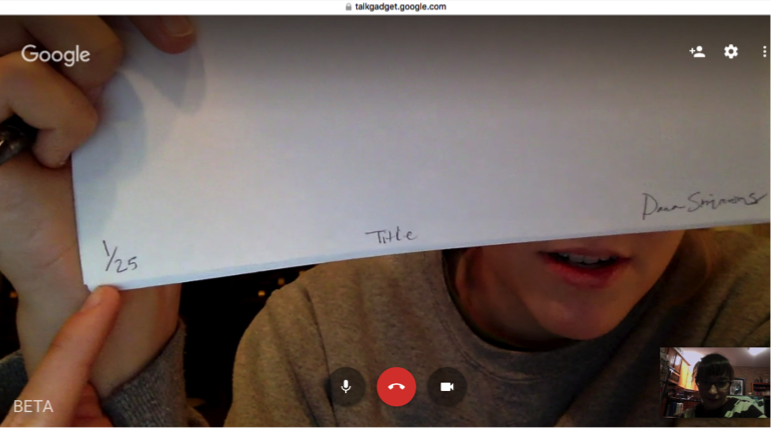
Here, Richelle showed me how to properly sign my name on a digital print, where to put the title, and where to put an edition number.
​My plan for this week is to print everything else I’ll need at the show, and to finalize the text for my collaborative project of illustrating science with Richelle.  In finalizing the text, I want to make sure that I didn’t write anything that’s scientifically incorrect while trying to write with non-technical metaphors.  Also, I may add a few new texts for Richelle to illustrate.  Specifically, I am thinking about debunking the myth of being “right brained” or “left brained.”  (In case you’re curious, you should know that your whole brain works all the time.  Unless you have a specific disease.)  I also would like to debunk the myth that people use 10% of their brains.  People are complex – we perform complex actions, use language, make coordinated movements, think about complicated stuff, and all these actions keep our whole brain working all the time.
 
Here are photos of some of the images I’m planning to bring:
 
Contact me ([email protected]) if you are interested in the art and want me to send you a photo where it’s easier to see the prints individually.  I’m thinking about starting a website to display my art more clearly, but for now, just email me and I’ll happily send you photos and details.
Picture
Picture
Picture
0 Comments

Dana & Richelle: Week 16

1/19/2016

4 Comments

 
Dana's update:

This week, I had seven of my neuron micrographs professionally printed.  I’ve been looking into where to get high quality prints and different types of paper.  After finding a place to make my prints, and selecting the brightest white paper, I sent in seven images for a test print.
 
When I returned a few hours later to pick up my prints, my face literally lit up with joy.  The colors were vibrant, the dendrites were huge, and I could see so many dendritic spines!  This was the first time I had seen my neurons printed so large and in color.  Normally, I only look at these images in the lab and on my iPhone screen.  The actual prints are a very exciting step up from my cell phone screen.
Picture
Here are the first seven prints of my neuron micrographs: three full cells and four close-up images of dendrites. The sizes are 12” x 12” and 12” x 18”.
Already, four people have asked me to make additional prints for them, which I will gladly do.  Two different people have expressed interest in hanging these in UChicago’s new neuroscience institute, which I would absolutely love.  The institute is brand new, so the walls are completely empty and very white.  I want these to be accessible so everyone can see what neurons look like.  I want everyone to be able to appreciate the intersections of science and art. 
 
My next step is to print these even bigger, add more images to the collection, and maybe think about printing them on sheets of metal.  I also need to explore how to hang/frame these for the upcoming symposium at the SciArt Center.  Richelle and I are meeting via Google+ on Tuesday night, and I’m looking forward to asking for her advice on how I should display my neurons.  Hope to see you at The Bridge symposium on February 17th.
 
Cheers!
Richelle's update:

For the symposium, I will have 10 new drawings completed.  These illustrations are visual interpretations of Dana’s writings on autism, neuroscience, genetics, and other ponderings about our humanity. This is a fun project for me to discover new imagery and subject matter.  Our collaboration continues to elaborate my own interests in connectivity, neurobiology, and evolution!
Picture
Drawings in progress.
4 Comments

Dana & Richelle: Week 15

1/12/2016

0 Comments

 
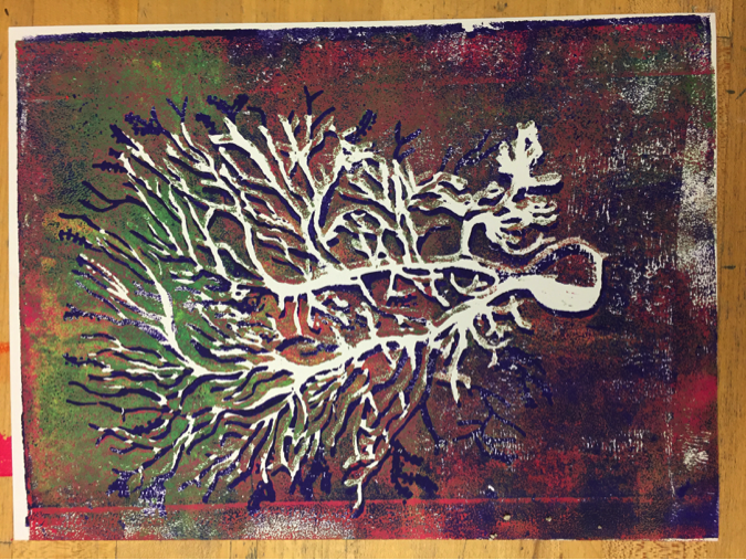
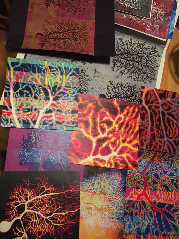
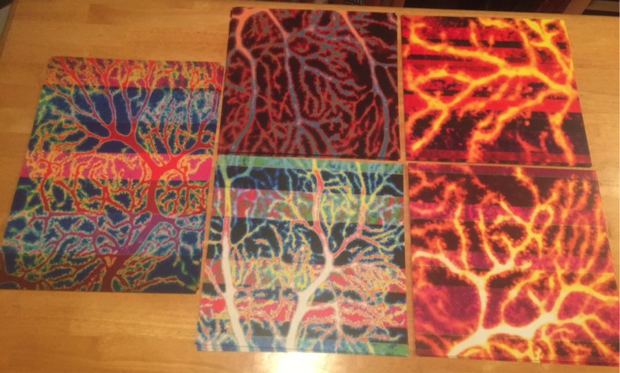
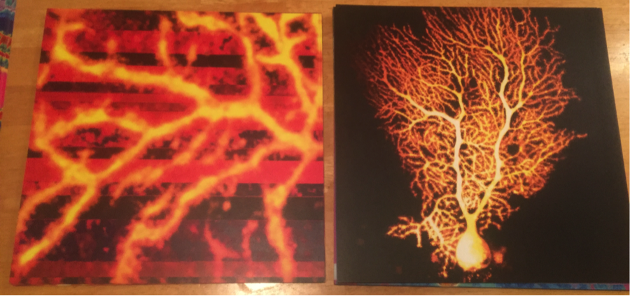
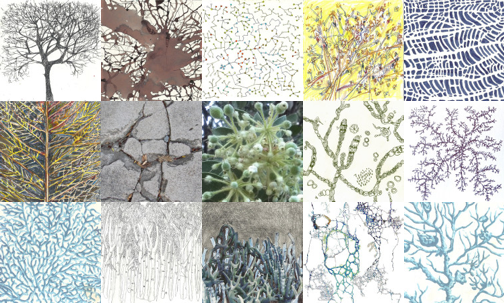
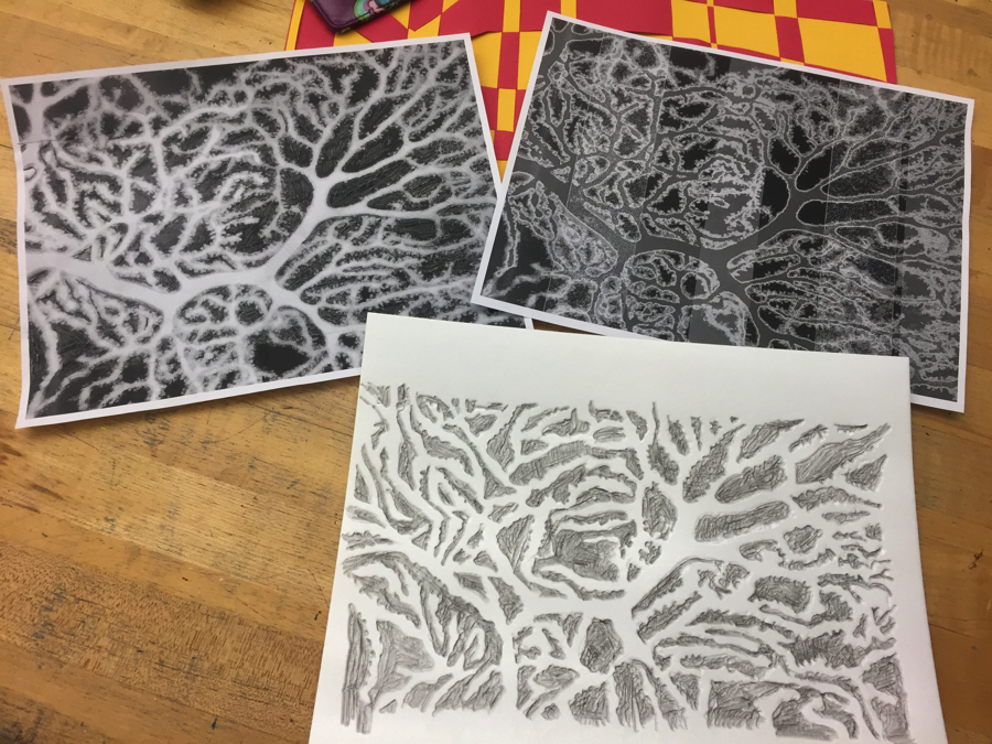
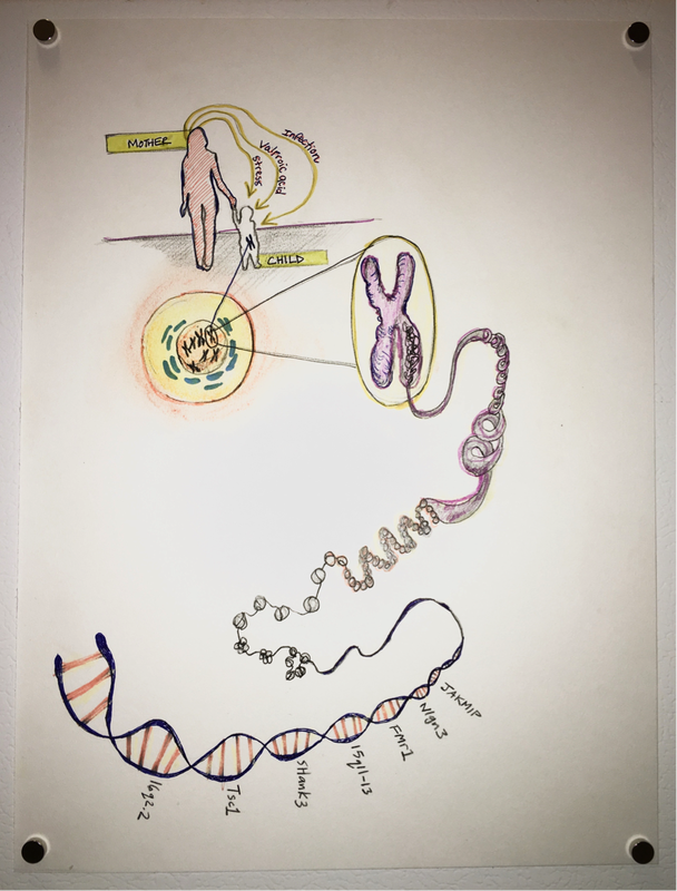
Richelle's update:

This week I found source material to brainstorm several new illustrations. Content includes stress symbols (sticky notes, flustered faces, clocks, tense muscles, etc.) in response to Dana’s question, “[i]s autism more common now than in the past?” As discussed in her response, environmental factors like stress on the mother may cause autism to be more prevalent in her child.  I am starting to merge various symbols of stress into a clock-like form to imply how a highly demanding busy life can cause symptoms of autism inherited by the youth. This is still being evaluated, but seems to be a topic of research and inquiry. Drawing coming soon. 
Picture
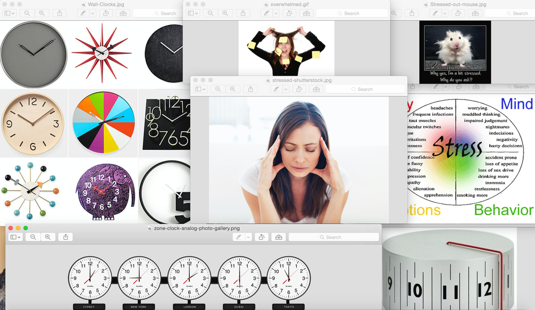
​In addition to continually brainstorming and creating new drawings, it is time to consider installation ideas for SciArt Bridge’s upcoming showcase event at the School for Visual Arts, New York. Tonight, I am researching good framing options to display the image/text content for our pieces. A stunning way to showcase drawings is to “float” the artworks in a frame to create a shadow, which makes the artwork appear to be more 3-dimensional.  By utilizing white frames (as opposed to colored frames, metal, or exposed wood), the delicate drawings will be more prominent with a simpler and subtle frame. This upcoming week, Dana and I will be discussing our plans for installing and presenting our collaboration at the exhibition in February.  
Picture
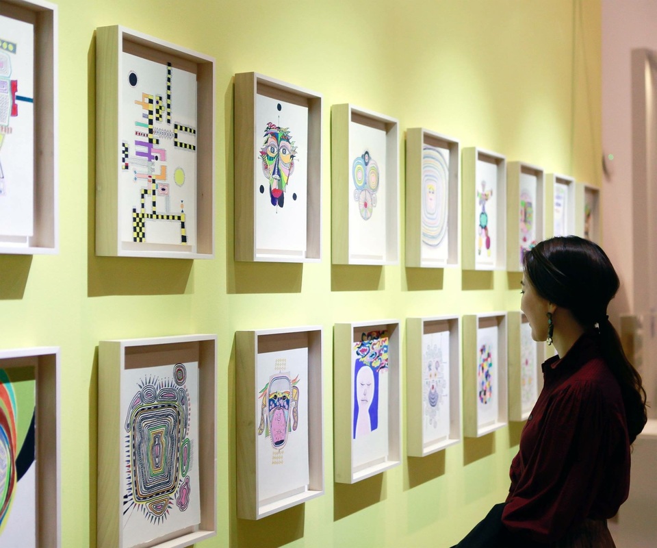
Brainstorming frames for use at the upcoming SciArt Center’s Bridge symposium and exhibition.
0 Comments

Dana & Richelle: Week 14

1/5/2016

1 Comment

 
Dana's update:

This week for The Bridge, I’ve been working on the text for my collaboration with Richelle.  I have written answers to three questions about Purkinje cells and finding Purkinje-like shapes elsewhere in the world.  I’m glad to be collaborating in this project because it gives me the opportunity to think more abstractly about my niche area of research and apply it to other things in nature. Each time I look for Purkinje patterns in the world, I find more and more examples.  Here’s what I’ve written on this so far:
 
“What are other examples of the Purkinje pattern in nature?
 
Purkinje cells exhibit the most complex dendritic branches of all neurons.  The dendrites of a Purkinje cell begin with one large branch that splits into two smaller branches, which further divide into medium sized branches, that eventually split into many, tiny, winding dendrites in an almost fractal nature.  What is so visually striking about a Purkinje cell is how closely it resembles the shape of a tree.  The tree shape above ground begins with a thick trunk that gradually divides into increasingly smaller branches.  This tree shape, or Purkinje pattern, is found both microscopically and macroscopically across nature. 
In addition to trees and neurons, the Purkinje pattern appears in nature in antlers, coral, blood vessels, river tributaries, and broccoli, just to name a few.  Even beyond nature, we have found virtual ways to use the Purkinje pattern in phone-tree networks, outlines with bullet points, and organizational file folders on computers.  We use this structure to add clarity and efficiency to our lives, and we can speculate that it formed in nature so many times for a similar reason.”
 
In addition to working on the text, I finished up the dendrite science-art that I was working on last week.  Here are some photos of my prints:
Picture
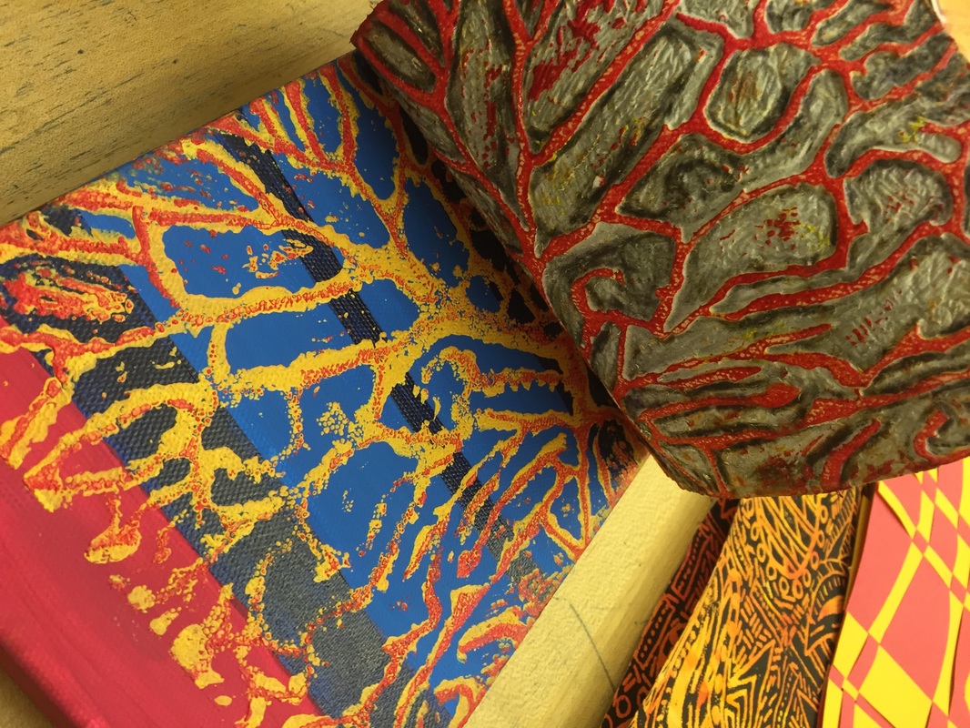
This image shows dendrites that I printed onto canvas in an effort to recreate one of my images from the microscope.
Picture
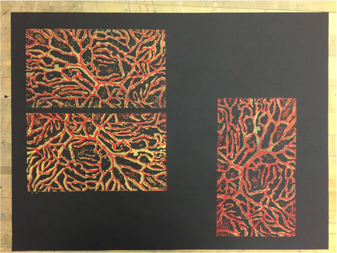
Here, I printed two layers of dendrites onto black paper. I haven’t decided if I want to cut out the rectangles or keep them all together on this paper yet.
Picture
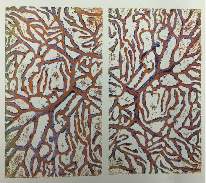
I also printed some dendrites on white paper.
​Check back next week for another update from me and Richelle!
1 Comment

Dana & Richelle: Week 14

12/30/2015

0 Comments

 
Dana's update:

In last week’s entry, I posted a list of questions that I plan to answer for my collaborative project with Richelle.  The first question I focused on this week was about finding Purkinje patterns in nature.  A Purkinje pattern is something that starts as one main channel and branches out many times, each time branching into smaller and smaller divisions.  I think one of the best examples of a Purkinje pattern in macroscopic nature is seen in tree branches. 
 
In the photo below, the tree trunk branches off into several main sections, then divides further into tiny branches.  Similarly, a Purkinje cell has a main dendrite that usually branches into two large dendrites.  The two large dendrites quickly split into tiny little branches, where most dendritic spines are found.  Dendritic spines look like little stubs that stick out from dendrites.  Often, when I see trees decorated with Christmas lights, the lights remind me of dendritic spines.
Picture
Can you recognize the pattern that resembles Purkinje cell dendrites in the two trees with Christmas lights in this photo? Note: Though the tree has lights around the trunk, it’s important to remember that Purkinje cells have most of their spines in the small dendritic branches, not the large primary branches. I took this picture yesterday in Chicago’s Federal Plaza. The red sculpture is Alexander Calder’s “Flamingo”, and the buildings in the plaza were designed by Ludwig Mies van der Rohe.
​For comparison, here’s one of my new photos of a Purkinje cell from the lab:
Picture
Here’s a new Purkinje cell photo I collected at the end of my experiments last week. Can you see the tree branch structure in this neuron?
​In addition to answering neuroscience questions, I’ve been working on printing some dendrites.  I first trace a printed image of some of my dendrites onto foam, creating a printing plate.  Then I will roll ink onto the template and press the foam into paper to print my dendrites.  I’ll post a photo next week with some of the prints after I finish making the foam plate. 
Picture
Here’s the foam printing plate I’m working on (bottom) along with two printed images of dendrites I imaged under the microscope.
​Check back next week for news about my collaboration with Richelle!
Richelle's update:

One of the drawings completed during the Bridge program is currently on display at Kala’s Artist’s Annual Exhibition at Kala Gallery in Berkeley, CA.
Picture
Opening reception of Kala Artist’s Annual at Kala Gallery on Thursday, Dec. 17, 2015.
​The selected drawing for exhibition is “Intertwined-4,” which combines various network forms including neural imagery contributed by Dana. It was exciting to share this work to more people and hear their input. Some gallery visitors suggested this drawing be enlarged into a giant mural or made into a puzzle to promote connectivity through interactions. Others were very interested in seeing the complete set of drawings because they are interrelated and visually link together.
Picture
Intertwined-4 is the second from the left on the wall.
YOU’RE INVITED!
 If you find yourself in the Bay Area in the coming months, take a look! 
Picture
​Kala Artists' Annual Exhibition
December 17, 2015 – March 26, 2016
Gallery Hours: Tue-Fri, 12-5:00pm; Sat, 12-4:30pm
Gallery location:
2990 San Pablo Avenue, Berkeley, CA 94702 
510-841-7000
0 Comments

Dana & Richelle: Week 13

12/22/2015

0 Comments

 
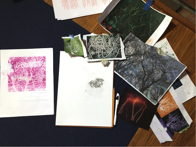
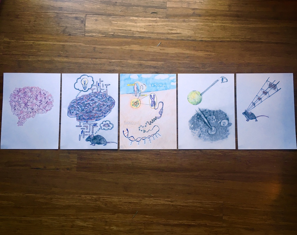
Dana's update:

This week, Richelle and I talked about more questions we could pose for our collaborative project, which will use both text and images to explore neuroscience.  So far, most of the questions have been about autism research and why scientists work with animals.  Richelle suggested that she could aim for more variety in the images if I wrote about more different topics.  Since we were originally matched to collaborate based on our interest in microscopic and macroscopic nature, we discussed posing some questions about these patterns.  With texts about what cerebellar Purkinje cells and other neurons look like, Richelle will have a chance to illustrate my neurons and draw comparisons between large Purkinje-shaped structures in macroscopic nature.  So far, Richelle and I have found similar structures in Purkinje cell dendrites, coral, social networks, river tributaries, trees branches, tree roots, and broccoli.
 
Here are some questions I am looking to create short texts for:

  1. What is a Purkinje cell?
  2. What do Purkinje cells look like?
  3. How are Purkinje cells different from other neurons?
  4. What are other examples of the Purkinje pattern in nature?
Picture
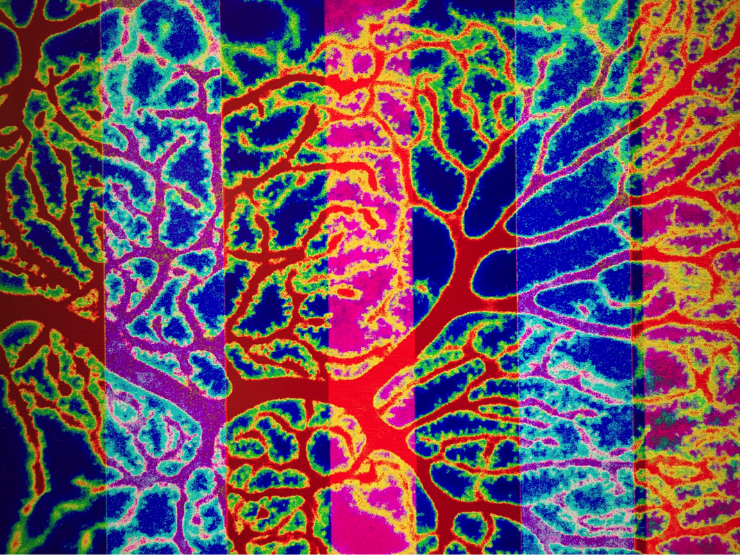
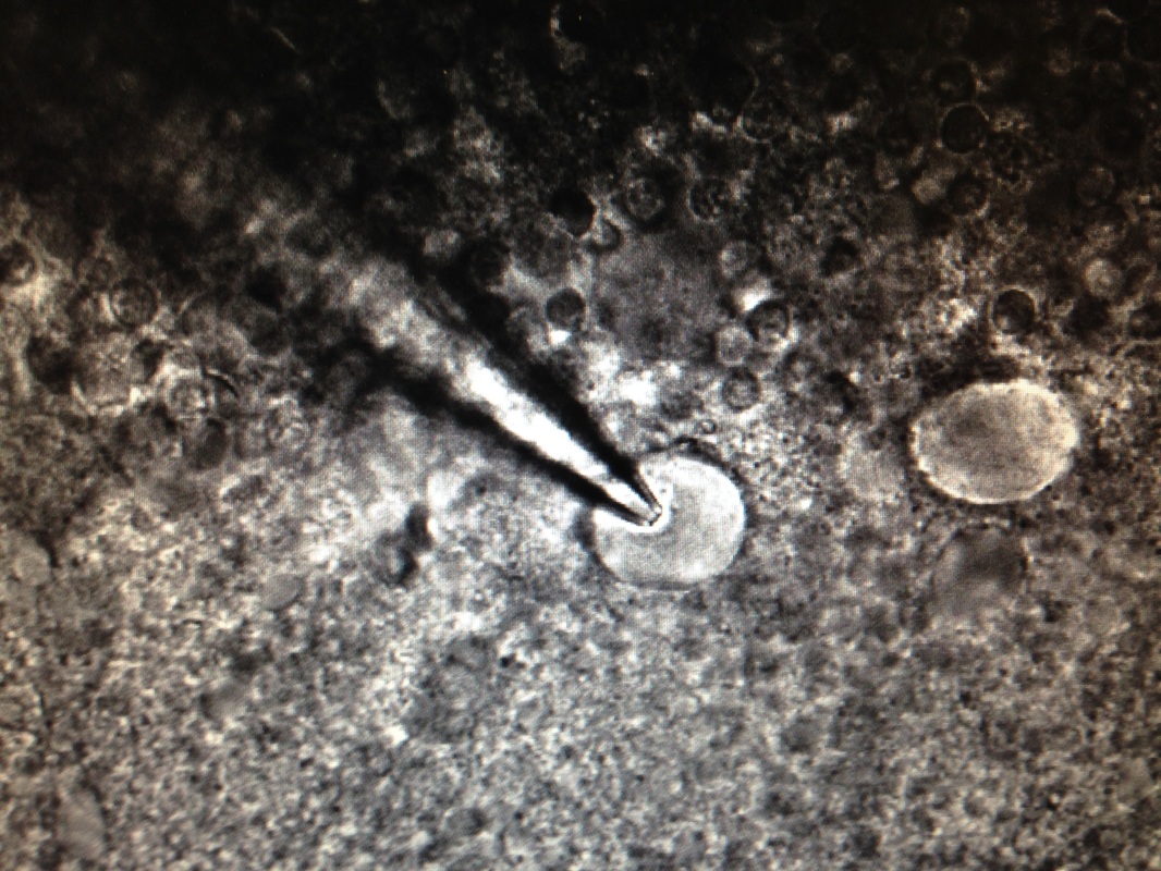
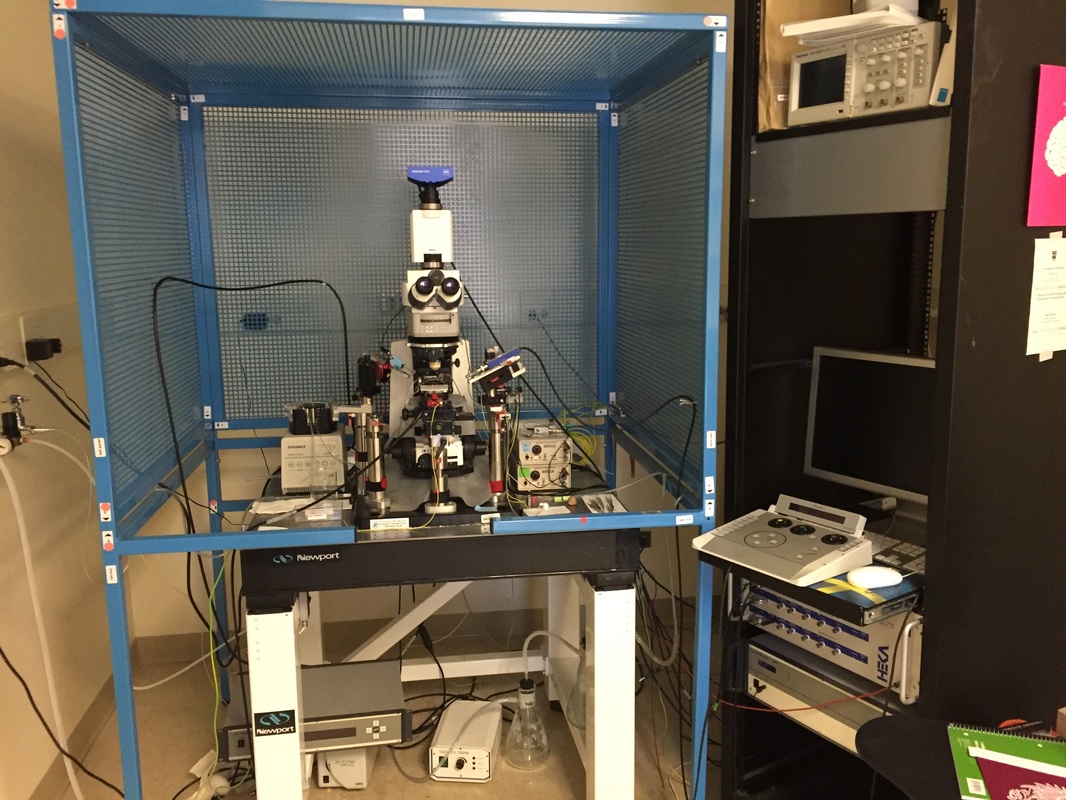
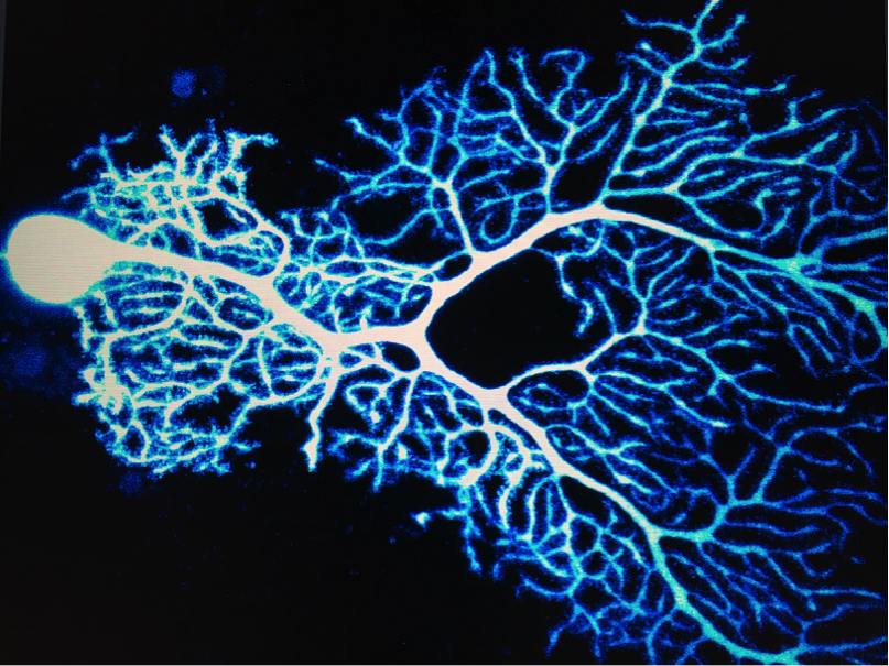
This is a photo of a Purkinje cell, which is a neuron in the cerebellum. I took this picture a few days ago in the lab by acquiring a z-stack of about 50 images in planes that were vertically separated by ~0.85 µm using our confocal microscope.
With these texts, Richelle and I would have some entries about autism, and some about cerebellar neurons.  I think it would also be great to create entries about experimental techniques such as patching cells, as well as to create entries about who scientists are today and what daily life is like as a scientist.  Maybe we’ll even create some entries about how to slice a brain!
 
Here are some other questions I would like to answer in this collaboration:
 
  1. What does a scientist look like today?
  2. What do dendrites do?
  3. What do neurons have to do with forming memories and learning?
  4. How do you slice a brain? 
Picture
Here’s a picture of some Purkinje cell dendrites that I used in a recent calcium imaging experiment.
​If you have questions about the brain, autism, or anything else in neuroscience, tweet them @dhsimmons1, and I’ll get you an answer.  Cheers!
0 Comments

Dana & Richelle: Week 12

12/15/2015

0 Comments

 
Dana’s update:
 
This week for The Bridge, I talked with Richelle about planning for the symposium at the SciArt Center in February.  Our goal is to get about 10-15 illustrations done to match with short texts about my neuroscience research.  In addition, Richelle has incorporated my neurons into some of her larger drawings that she might bring, and I have a few photos of neurons I would like to bring.  Richelle recommended that I try to print some of my best neurons on aluminum sheets to bring out the colors.  At the moment, I’m looking for a printer in Chicago who can do something like that, because I think the idea is super cool.
 
My next text that Richelle is planning to work with is about patching neurons.  In my paragraph, I explain the technique of patching, and liken it to sticking a straw into a tennis ball.  (Basically, when you patch a cell, you stick a tiny glass tube into a neuron.)  In order to provide Richelle with all the imagery she’ll need to illustrate the concept, I’ll be sending her more photos from the lab.  Yesterday, I emailed two photos to Richelle.  The first photo shows a class tube (pipette) extending from the cell body of a Purkinje neuron, which is the patched cell.
Picture
This image is from a light microscope that projects to a camera. In the middle, you can see two round shapes, which are the cell bodies of two different Purkinje neurons. The cell body in the middle has a glass tube (pipette) pushing into its center in order to patch the cell. When the cell is patched, the fluid inside the glass tube can flow freely into the fluid that’s inside the cell body. Making this type of link between the tube and the cell allows me to fill cells with dye for imaging experiments.
The second photo is a larger view of the microscope I use to patch neurons.  
Picture
In this image, you can see the microscope in the middle, surrounded by a blue Faraday Cage. The Faraday Cage helps reduce noise from air vibrations and movements in our recordings. The microscope floats on a table that is similar to an air hockey table to further reduce vibrations. On the right, there’s a computer I use to monitor the cell’s electrical activity once it’s patched.
​Patching cells is an exquisitely delicate technique, so we have to be very careful not to let the microscope move.  If you so much as flick the microscope lightly with your finger, the patch between the pipette and the Purkinje neuron’s membrane will immediately break, and you’ll have to start over with a new neuron.  In order to reduce air vibrations, the microscope is surrounded by a blue box called a Faraday Cage.  To reduce the effects of experiment-ruining building vibrations, we place our microscopes on what is sort of like an air hockey table.  As long as nothing moves, we are able to keep a 2-µm pipette precisely centered over the 15-µm cell body. 
 
In other news, I am considering making a series of food sculptures shaped like Purkinje cells.  I’ve been compiling a list of food items that would make good cell bodies, dendritic spines, dendrites, etc.  This project is still in the brainstorming stage, but I am hoping to build a few of these soon and photograph them.  Wouldn’t it be cool to see a Reese’s peanut butter cup as a cell body with Pull ‘n’ Peel licorice as dendrites and Mini M&M dendritic spines? 
 
Check back next week for a new update on my collaboration with Richelle!
Richelle's update:

Question + Artwork Below…

From Dana: 
What does it mean to “patch” a cell?
Patching a cell is the microscopic equivalent of sticking a straw into the side of a tennis ball, only much more delicate.  Once your straw is attached to the tennis ball, you’re free to look through it and see what’s going on inside the tennis ball.  In the microscopic world, the straw is a tiny glass tube called a pipette, and the tennis ball is the cell body of a neuron.  Researchers use microscopes and micromanipulators to place a pipette delicately so that it gently presses into the cell body of a neuron that is about 20 µm wide.  Once the pipette is pressing on the cell, a researcher can apply tiny amounts of negative pressure to suck the cell membrane up against the opening of the pipette.  With just the right amount of negative pressure (basically sucking on a straw gently), a researcher can open the portion of the membrane that’s inside the glass pipette opening while leaving the rest of the neuron intact.  Once the membrane is open to the pipette, the neuron is considered “patched”. 

The reason scientists patch cells, or use the patch clamp technique, is to listen in on a neuron’s activity.  Using patch clamp methods to record a neuron’s electrical activity is like eavesdropping on a conversation happening next to you at lunch.  Through a glass pipette, researchers can monitor how a neuron communicates with the cells around it.  Neurons communicate using electrical signals as their language, and they respond to electrical signals from nearby neurons and cells.  We can monitor these conversations to learn about the physiology of different types of cells.

In my research, I patch neurons called Purkinje cells in brain slices from the cerebellum of autistic mice.  My goal is to look for differences in the electrical activity of Purkinje cells and their surrounding cells between the autistic and non-autistic mice.  By finding differences in electrical conversations in the brain slices from autistic mice, I hope to determine what is different in the autistic brain.
My artwork in response:
Picture
Drawing for “What does it mean to ‘patch’ a cell?” – artwork is in progress.
0 Comments

Dana & Richelle: Week 10&11

12/8/2015

0 Comments

 
Dana's update: 

This week, I’ve been collecting photos around the lab to send to Richelle so that she has an idea of what to draw for our collaboration.  I’m mostly sending photos of bottles, beakers, pipettes, and things around the lab benches.  Seeing Richelle’s drawing from last week was so exciting!  I’m amazed by how much science she incorporated into the image, and I love that both the human brain and a mouse brain are represented in the artwork.  Can’t wait to see the next set of images!
 
Here are some photos that I sent Richelle so she can see what the lab looks like:
Picture
This is one of our lab benches. In the foreground, we have reusable Styrofoam containers that we use to ship brains to collaborators. When we’re not mailing brains, we use these containers as ice buckets to keep things cold. We also have lots of little bottles on the shelves that hold various reagents that we use in experiments.
Picture
This photo shows pink and yellow racks for holding tubes. We mix small amounts of liquids in these tubes. The shorter yellow rack is sized to hold 1.5 mL tubes. The pink and large yellow racks hold 50 mL conical tubes. We have a pH meter sticking out of the open tube in the tall yellow rack.
Picture
The plate on the left is a magnetic stirrer. The flask on top of the plate has liquid and a magnetic stir bar in it. When you turn on the plate and choose a speed, the stir bar spins and mixes the liquid. In the middle, we have a standard scale. On the right, we have a more sensitive scale with doors on each side so that fluctuations in air currents won’t affect the measurement.
​I’m hoping that Richelle is finding the photos and captions helpful in creating her next artworks, and I’m very much looking forward to seeing her drawings!  Check back next week for another update.

Richelle's update:

I am in the process of interpreting Dana’s text questions into visuals. I realize that as I make new drawings, my impulse is to add to other drawings. This process creates a more cohesive aesthetic among all pages of the science-art book project.  The drawings below are not yet complete and are (potentially) the first layer of what will become more complex drawings. I anticipate I will add bolder colors, thicker lines, and more texture as these drawings evolve.
 
Question + Artwork Below…
 
Why do scientists use mice to study diseases?
 
What causes autism?
The current scientific consensus is that autism is caused by both genetic mutations and some environmental factors.  Genetics play the main role in causing autism.  Many different genes have been identified as being associated with symptoms of autism, but researchers still cannot zero in on one causal gene.  In light of recent autism research, many researchers who study autism now speculate that autism is the product of mutations in many different genes that work together.  Since there are many types of autism (it’s a spectrum disorder), it seems plausible that there are different mutations in different genes that are responsible for each type of autism.  When these genes are identified as mutated in autistic patients, researchers often create mice that model the mutation in order to study the gene.  Some genes scientists are currently studying for their association with autism include: JAKMIP, Nlgn3, Fmr1, 15q11-13, Shank3, Tsc1, 16q2.2, and many more.
In addition to genetics, many researchers believe autism can be caused by certain environmental factors. Environmental factors that researchers have connected to autism are based on the health of the mother.  Infection, valproic acid (found in some medicines), and stress level of the mother during pregnancy may have an effect on whether or not the child will have autism.  Numerous studies have concluded that environmental factors such as pesticides, GMOs, and vaccines do not cause autism. 
Picture
Artwork not yet complete! I want to further emphasize how autism is caused by BOTH genetic mutations and the health of the mother (infection, valproic acid, and stress levels). I will somehow divide this page to indicate internal vs. external influences that generate autism.
​Other drawings posted below have a similar aesthetic. For these drawings I am using graphite, charcoal, colored pencil, and ink. I may add in some watercolor paint as well. Finally, the drawings may even be scanned and manipulated graphically like the previous blog post. As you can see, I enjoy working on multiple artworks at one time and making changes as I go along!
Picture
Drawing for “How do mice show symptoms of autism?”
Picture
Drawing for “Why do scientists use mice to study diseases?”
0 Comments
<<Previous

    GROUP TWO

    Picture
    Dana Simmons
    Picture
    Richelle Gribble

    RSS Feed

Powered by Create your own unique website with customizable templates.