SciArt Initiative

  • Mission
  • Program Archive
    • Events
    • Exhibits
    • Residency
    • Colloquium
    • Magazine
  • Mission
  • Program Archive
    • Events
    • Exhibits
    • Residency
    • Colloquium
    • Magazine

Dana & Richelle: Week 9

11/24/2015

0 Comments

 
Dana's update:

This week for The Bridge, I was thinking about the complex branching structure in Purkinje cell dendrites.  Purkinje cells look very different from other cells because of their enormous dendrites. Neurons are often classified by their shape. I want to use this week’s post to emphasize how a Purkinje cell’s shape stands out from other cell types. I sent Richelle some photos of other types of cells, and I’d like to share those photos here with you.
 
For reference, let’s start with a Purkinje cell. Here’s the latest Purkinje portrait from my experiments last week.  Look at all those dendrites!
Picture
In this photo, you can see the Purkinje cell’s cell body (round part at the bottom) and the large dendrites extending up from it like a tree. You can even see some of the dendritic spines (little dots on the branches). Dendritic spines are the molecular sites where learning and memory happen.
​The next cell type of cell I want to show you is called a glial cell (singular = glial cell, plural = glia / glial cells).  There are many types of glial cells. Glial cells are not neurons; rather, their purpose is to nourish and support neurons, and make electrical impulses travel faster through neurons. Glia are found all over the nervous system, both inside and outside the brain. Here are some glial cells that I photographed as an undergraduate researcher at Boston University back in 2010.
Picture
In this image, you can see the outline of the glial cell in green. This cell was stained with green fluorescent protein (GFP). The blue part (stained with DAPI) is the nucleus, which is where the cell’s DNA resides.
Picture
Here’s another glial cell. As you can see, glia come in all shapes and sizes. The shapes of the glial cells in these photos vary a lot because these cells were not photographed while in brain slices. These cells lived in petri dishes and on small cover slips, where neurons and glia were cultured together, and then placed onto slides for viewing.
​In the next photo, you can see both glial cells and neurons from a part of the brain called the hippocampus. The hippocampus is generally responsible for memory, and it’s a hotspot for research on Alzheimer’s disease.
Picture
There’s a lot going on in this photo. The spread-out glial cells from the previous two photos were from the edge of the culture in the petri dish. In contrast, this photo came from the middle of the petri dish where things are more crowded. The blue dots (kind of look like grapes) surrounded by green are a crowd of glial cells. Above those, you can see hippocampal neurons called pyramidal neurons. Pyramidal neurons have smaller nuclei than these glia, and they have green extensions that are marked with red dots. The red dots are from a stain that allows us to see which of the green extensions are dendrites, and which one is the axon. Since the red marks dendrites, it will not be present on the axon. In a brain slice, it’s easier to know which extensions are dendrites and which is the axon; however, in a cell culture in a petri dish, all the extensions from the cells go in every direction and get all mixed together. We use special stains to tell them apart.
Now that you know how to recognize Purkinje cells, pyramidal cells, and glia, I want to show you one more image to emphasize the differences between photographing a neuron in a brain slice vs. a petri dish. In the previous photos, the pyramidal cells’ extensions go in any direction they want because the neural circuit they came from is disconnected. This next neuron is also a pyramidal cell, however, the photo was taken in a slice (and using completely different experimental techniques).  In the slice, the pyramidal cell grows its dendrites and axon in the right direction, toward the other brain structures with which it communicates. The photo below shows a pyramidal neuron in a slice. The neuron is stained with a dye, which clearly shows the cell body (middle dark spot) and begins to travel down the axon and dendrites.  This neuron beautifully shows the characteristic triangular shape of a pyramidal cell in the brain (not a petri dish), and was photographed by Heather Titley, a Postdoctoral Scholar at The University of Chicago. Heather and I work together in the Hansel Lab.
Picture
This image was taken by Heather Titley, a Postdoctoral Scholar at The University of Chicago. The picture shows a pyramidal neuron (See the characteristic triangle shape?) from a frontal region of the brain called somatosensory cortex.
​Neurons (and all cells) come in many different, beautiful, and fascinating shapes and sizes. If you want to see more types of cells, look up “starburst amacrine cells” and “menorah cells” on Google images. These days, neuroscientists are using neuronal morphology to classify neurons. Neurons are typically classified in many ways, but there’s no reigning consensus on whether it’s best to classify by shape, size, location, what neurotransmitters they use, or something else. Regardless of how you prefer to classify neurons, I believe we can all appreciate the art that results from imaging different types of neurons around the brain.
 
Here’s a link for photos of menorah cells: http://www.livescience.com/40321-worms-tell-tale-of-how-nerves-develop.html
 
Here’s a link for photos of starburst amacrine cells: https://www.google.com/search?q=starburst+amacrine+cells&client=safari&sa=X&rls=en&biw=1274&bih=652&tbm=isch&tbo=u&source=univ&ved=0ahUKEwjk6LO2_KHJAhWFSiYKHZlMBAgQsAQINA
 
Check back next week for another update.  Cheers!
Richelle's update:

This week I created some illustrations in response to a text question/answer written by Dana. I am experimenting with styles right now. I created several sketches to brainstorm content and now I am rearranging this material to create experimental graphics. 
Picture
Compiling images of the brain/mouse maze, thought bubbles, human brain, mouse brain, light bulb. New ideas come to light by researching mice to understand our humanity.
Question + Artwork Below…
 
Why do scientists use mice to study diseases?
 

In research, it is increasingly common to use mice to model diseases. Today, mice can show symptoms of autism, Alzheimer’s, Parkinson’s, Schizophrenia, ataxia, cancers, and almost any other disease and disorder you can think of. Using mouse models of diseases and disorders enables researchers to learn about how the condition hurts the brain, and attempt to devise treatment strategies. The overarching goal with translational rodent research is to see what went wrong in the brain, and try to fix it with therapies that could potentially alleviate suffering for people who have these diseases and disorders.
 
Most researchers would prefer to use a model of the brain and stop bugging all these mice. However, creating a model brain assumes that we already know everything about how the brain works. In order to create a useful model, researchers would truly have to understand how every part of the brain functions and connects to other parts. Despite huge advances in neuroscience, we’re quite a long way from knowing this much about the brain.  Until we have such a deep knowledge, we must rely on mice or other model organisms to model diseases for us.
 
Historically, there have been cases where research animals were obtained inappropriately, and their well-being was ignored. Today, special regulations and committees are in place at research institutions to advocate for the safety and comfort of research animals.  These committees include experts in the field, veterinarians, and non-scientists from the community.  Scientists are prohibited from performing experiments until they have the committee’s approval. When deciding if it is appropriate to use animals in research, this committee considers ways to minimize stress and discomfort for the animals, as well as the importance of the research. In all research, these committee members and the scientists conducting the studies take care to demonstrate respect for research animals. 
Picture
Artwork 1: Why do scientists use mice to study diseases?
0 Comments

Dana & Richelle: Week 8

11/18/2015

0 Comments

 
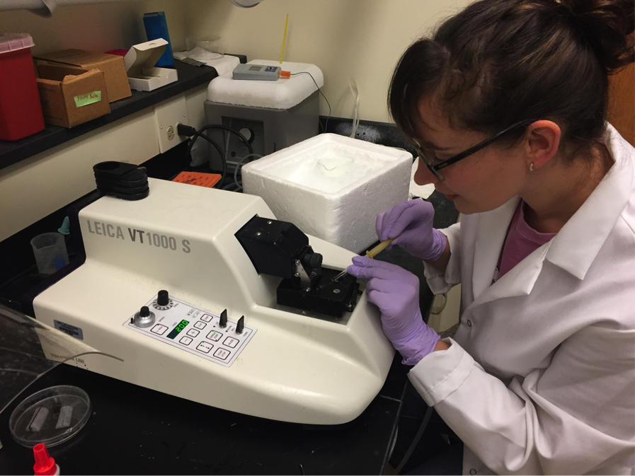
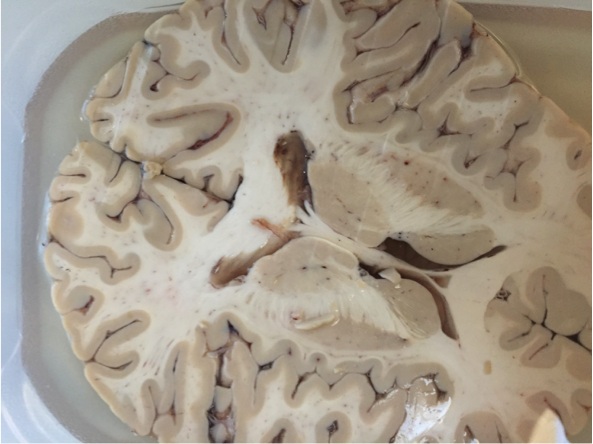
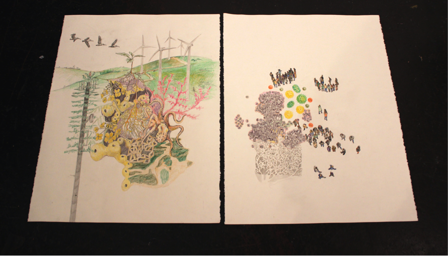
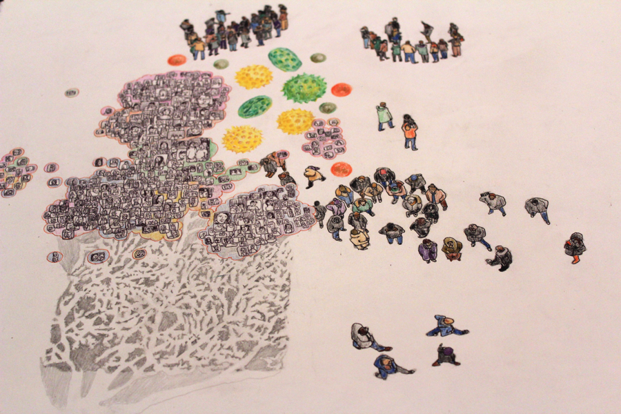
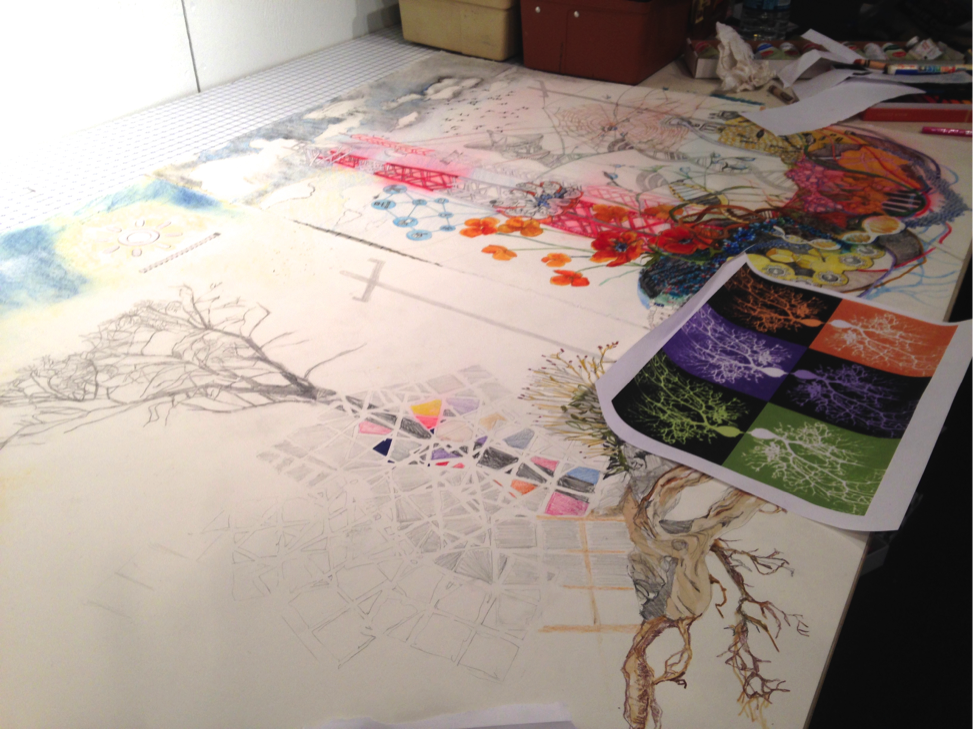
Dana's Update:

This week, in our biweekly meeting for The Bridge, Richelle and I talked about how neuroscientists slice brains for physiology experiments in labs.  Richelle had a lot of great questions about how we slice brains, what the equipment looks like, how we keep the neurons alive, and how long the process takes.  After chatting with Richelle, I decided to take some photos of the dissecting and slicing equipment and put together some captions.  My hope is that these images will be useful for Richelle when she creates images to go with my text for our collaborative project.  Here are some of the photos and captions that I sent Richelle:
Picture
In this photo, I’m placing the middle of the cerebellum (the tiny bit of brain tissue between the two dissection tools) onto a spot of superglue in the black box. Once the tissue is glued down, I fill the black box with a solution and use the blade (to the right of the tissue) to make 200 micrometer-thick live brain slices.
Picture
Here, you can see the cerebellum glued onto the platform just before getting sliced. The chunk of tissue is about the size of an eraser on a pencil.
Picture
Here, you can see the blade going through the tissue and cutting a very thin slice, which ends up on top of the blade. It’s a little blurry because the slicer is vibrating side to side as it gradually moves forward through the tissue. It takes about 20 seconds for the blade to make it all the way through the tissue for each slice. The dissection and all the slices have to be completed in about 12-15 minutes in order for the slices to be healthy enough to last through an experiment. We make slices in the mornings, and use the slices for experiments in the afternoons/evenings.
Picture
In this photo, I’m using a small suction tube to suck up a brain slice after the blade has completely disconnected it from the rest of the tissue. The blade starts at the top, and then moves down 200 micrometers for each slice to make sure they’re all the same thickness.
Picture
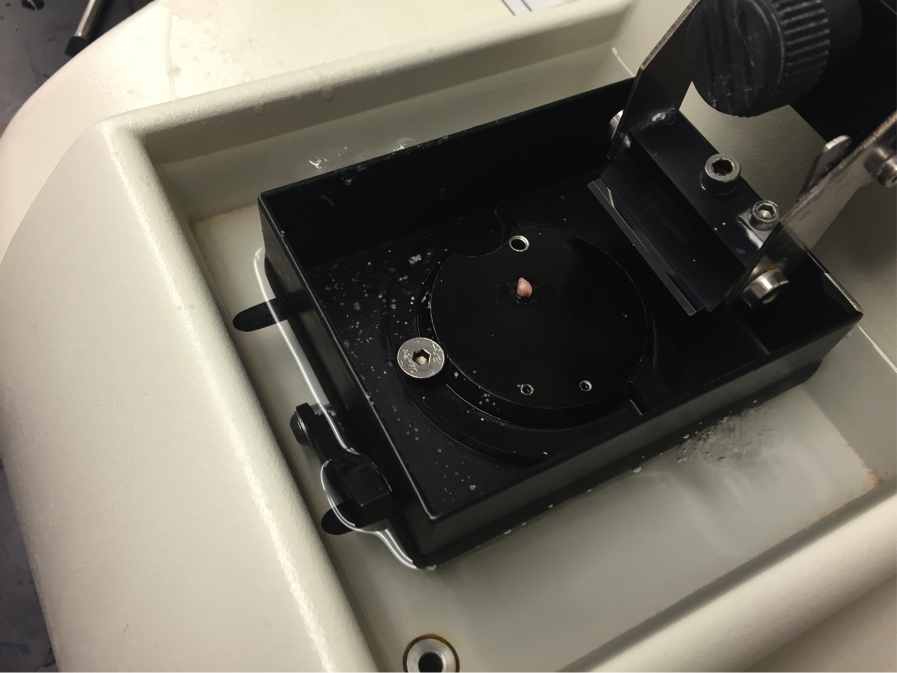
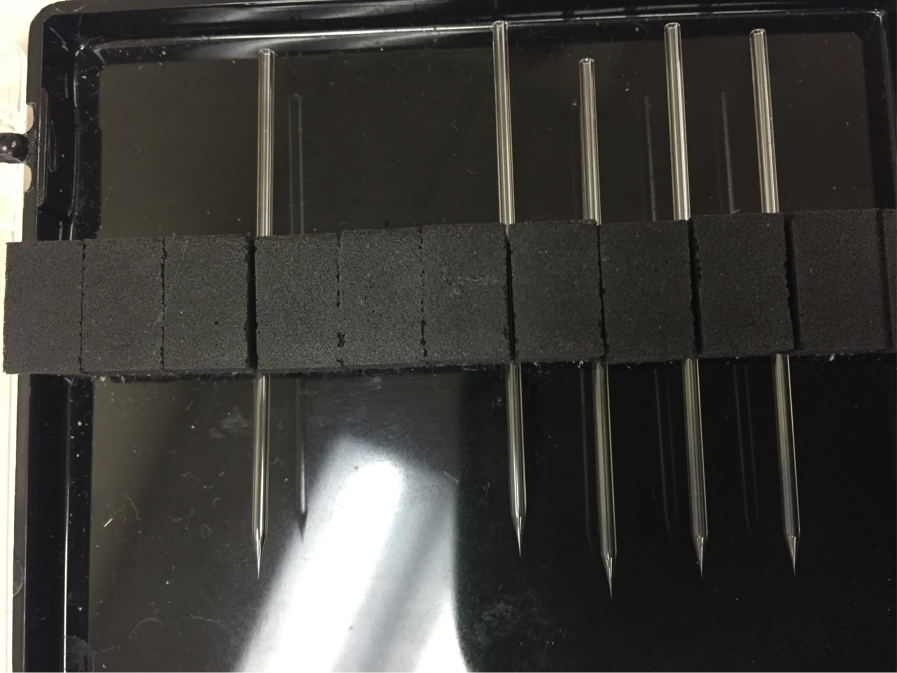
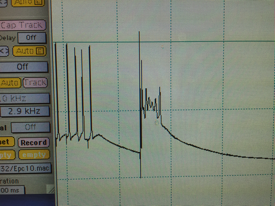
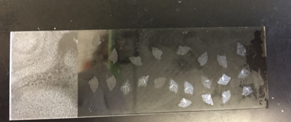
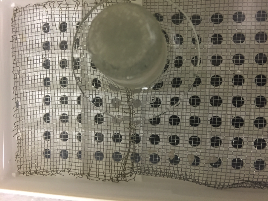
This is the chamber that the brain slices recover in for about 1.5 hours after getting sliced. If you look carefully in the bottom right corner of the photo, you can see three slices lined up on top of the silver netting. The slices incubate here inside this box with a solution that has nutrients to keep them alive. In order to stay alive, the neurons on these slices also require oxygen, so we have gas tanks that bubble oxygen into the chamber.
​In this video, you can see the blade coming across the brain tissue and generating a 200 µm slice of the cerebellum. 
 
In addition to chatting about how to slice brains, Richelle and I also talked about the pipettes I use.  These pipettes are special, glass micropipettes that we make each morning.  We attach these micropipettes to the membrane of a neuron in order to measure the electrical activity going on inside that neuron.  We also use these micropipettes to fill some neurons with dye for experiments that generate the colorful images I’ve been posting each week.  Here’s some of the pipettes info I send Richelle:
Picture
Here’s my pipette box. These pipettes (five here) are the little glass tubes that I attach to the membrane of a neuron to “patch” the cell and fill it with dye while recording the cell’s electrical activity. In a good day, I’ll use three or four pipettes – one or two to patch neurons, and two to send electrical stimulation into the neurons. In a bad day, when these break (they’re really small and fragile), I’ll go through ten or more pipettes. We make new pipettes each morning. Old ones are used to send electrical stimulation into neurons, and new ones are used to patch cells and fill them with dye.
Picture
Here’s an even closer view of one pipette. The pointy tip is open on the end, and the opening is about 2-5 micrometers in diameter. The portion of a neuron that I patch with this is about 17-20 micrometers in diameter. I actually held my iPhone up to the eyepiece on a microscope to take this photo. ☺ The black bar on the bottom is platinum wire that heats up and to fire polish the sharp glass edges of the pipette so they’re not so sharp. This helps ensure that the pipettes won’t tear the cell membrane.
Picture
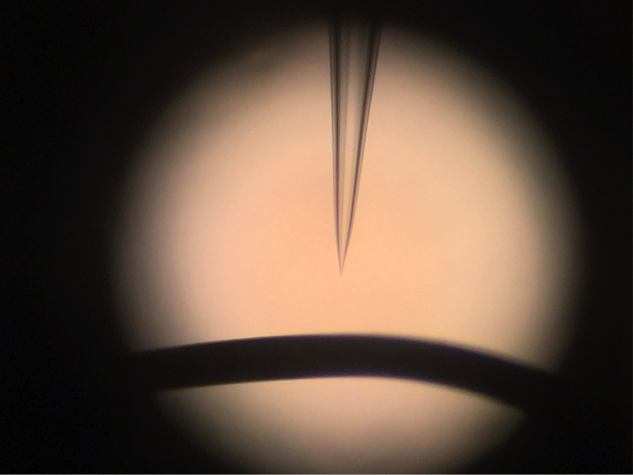
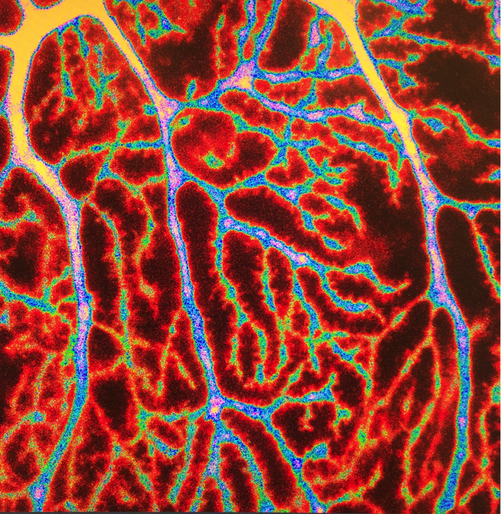
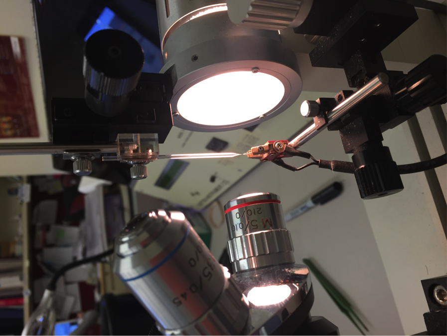
This photo is sideways….tilt your head 90º to the left. Here, I placed a pipette over the tiny platinum wire to fire polish it. The large lenses are attached to a microscope and help us see how the shape of the pipette tip changes as we fire polish it.
​Check back next week for another update.  Cheers!
0 Comments

Dana & Richelle: Week 7

11/10/2015

0 Comments

 
Dana's Update:

​​This week, I sent Richelle some short texts about neuroscience, autism, the cerebellum, and experimental techniques used in my research. We met on Google Hangout on Sunday to discuss our plans for the collaborative project, which will include both text and images. In addition to the texts I sent, Richelle suggested some additional questions that would be interesting topics to write about. In the next week, I’ll explore writing about differences between human brains and mouse brains, how scientists slice brains for experiments, and how mice can model diseases such as autism. 
 
In case you’re curious, this is what a mouse brain looks like:
Picture
This photo shows a mouse brain. The two large frontal hemispheres are on the left, and the cerebellum (the part I study) is on the right. A mouse brain is about the size of your thumbnail. In a mouse brain, the frontal hemispheres are smooth, but in a human brain, the frontal hemispheres are wrinkly and folded to maximize surface area for higher functions.
We’re thinking that we could hopefully continue our collaboration after The Bridge residency ends to learn more from each other and merge artwork with science texts. Both Richelle and I are excited about the future possibility of continuing our collaborative project and inviting more scientists to join the partnership to write about their own area of expertise. Maybe someday we’ll make a coffee table book!

​In addition to working on more text for our collaborative project, I’ve been working on some new experiments in the lab. I’m taking a few weeks away from my autism research to work on an experiment that aims to clarify how processes such as learning and memory happen in the cerebellum. For this new experiment, I’m still working with cerebellar slices filling Purkinje cells with dye. I use this dye to visualize how calcium travels through a Purkinje cell.  
Picture
This image (above) shows a page of my lab notes from the new experiment. I make notes about the health of the neuron, the quality of the patch between the glass tube (pipette) and the neuron, and also the types of electrical stimulation that I’m testing on that particular neuron.
Richelle's Update:

After meeting with Dana this week, I got to learn more about her day-to-day life in the lab.  As an outsider looking in, I asked simple questions to help visualize her experience. Do you work with a piece of brain everyday? What kind of mice do you study? What color are they? How do you know if a mouse is autistic? How big is the team at the lab? Is the brain alive when you work? All of these questions gave me a clearer idea of her routine, what the lab looks like, and how she works. Dana’s research is so precise, requires a quick hand, and is an intricate endeavor.
 
This week I reviewed several texts she sent to me that answer a range of questions, some specific to her research about autism and the cerebellum, and other broad questions that many of us (not wearing lab coats) have always wondered, like “why do scientists use mice to study diseases?” I decided to create a new drawing inspired by her answer to this question.  So far, I have compiled many images including the black mice (which are used at the lab), mice brains, human brains, mazes, modular forms, and more. This serves as a mood board for our text/art project.  A mood board typically consists of an arrangement of images, materials, or text intended to evoke or project a particular style or concept.    ​
Picture
My mood board - a collection of images/symbols that came to mind when reading Dana’s texts.
​For this drawing I hope to link the “man” and the “mouse” by overlaying imagery to show that they have striking commonalities. Dana’s text describes how mice are receptive to most human diseases and respond in similar ways, which is crucial for researching cures and understanding threats. I will post her final text after artwork is completed for her elaborate response. In this drawing, I hope to link imagery by creating a maze-like brain that ties the content together. It is fairly challenging to describe before I make it, but I have an idea that I am excited to show you when it is complete. I tend to work in layers of overlapping imagery so this drawing will most likely undergo a lot of changes. Below are the beginnings of this new visual interpretation of Dana’s question.
 
I look forward to turning her words to life! 
Picture
Contemplating ways to merge the iconic “mouse in a maze” symbol to link human brain and mouse brain in a seamless way.

Calcium plays a large role in the molecular mechanisms for learning and memory, so it’s important to be able to visualize where and when it flows through neurons. My goal with this experiment is to visualize where the calcium flows in response to different types of electrical stimulation. The different types of electrical stimulation mimic how we think the cells learn and form memories in coordination with the other nearby neurons.
​The data that I’m collecting with the new dye look a little different from what I collected with the old dye. However, from an aesthetic perspective, the neurons still look the same since all the color is added with imaging software. Without any dye, neurons are colorless. The normal color of a brain is a pale pink-ish hue that has some gray and some white mixed in.  Here’s a photo of a Purkinje cell that I filled with the new dye:
Picture
This image shows a recording for electrical signals in a Purkinje cell from my new experiment. The vertical fluctuations in the black line represent changes in the direction that ions (ex: sodium, potassium, calcium) are flowing in and out of a patched Purkinje cell.
​As a scientist, I’m not accustomed to doing things like transporting art around the county.  During our meeting, I asked Richelle for her advice on shipping art for The Bridge symposium in February. She told me about this cool way to make artwork on paper appear to be floating on the wall by using nails and magnets.  Since none of the work I’ve done is framed, I’m planning to look into that and try to float some of my art.
 
Have a great week!  Here are some more dendrites:
Picture
Picture
0 Comments

Dana & Richelle: Week 6

11/3/2015

0 Comments

 
Dana's Update:

This week for The Bridge, I am working on the series of prints that Richelle and I are creating. My part is to write a series of questions about autism and the cerebellum, and then provide answers in a couple of short paragraphs. I can’t wait to see the images Richelle creates to bring my text to life! Our goal is to add one entry per week for the rest of The Bridge residency, meaning that in total we’ll have about fifteen entries at the end. So far I’m up to twelve questions and five answers.
 
I’m excited to see how these prints turn out, and looking forward to seeing them both as an e-magazine and limited run print edition. If you have a question about autism and/or the cerebellum, tweet it to me @dhsimmons1.
 
Anyway, this week, I tackled questions about whether or not there’s more autism than there used to be and what causes autism. Let me start by stating that vaccines absolutely do not cause autism. Not even a little bit.  Numerous studies (go Google them) have shown that there’s no connection between autism and vaccines. In fact, the original paper linking them has been retracted by the journal (The Lancet), and the author of the study has been discredited. You can still look up the original paper, but now it has large red letters over the text that reads, “retracted”.
 
In between preparing some text for these questions, I’ve been spending a lot of time in the lab doing experiments to examine the physiology of the cerebellum in autism. I took a few more photos of really nice Purkinje cells (neurons in the cerebellum) and some dendrites. Enjoy!
Picture
Image: Dendrites on a Purkinje cell from the cerebellum of an autistic mouse. I added false coloring to the image using calcium imaging Zen 2008 software. In nature, dendrites, and all neural tissue, have a pale pink or white-gray-ish color. In this image, the red/orange “glow” around the branches is where the dendritic spines stick out. These spines are where important processes such as learning and memory happen at a molecular level.
Picture
Image: This is a photo of a Purkinje cell that is filled with dye. Near the upper left corner (circled), you can see a light cone shape, which is actually the shadow of a glass pipette with an electrode in it. During the experiment, I use this electrode to deliver small electrical pulses to the Purkinje cell, and I measure how the cell responds in order to learn about how Purkinje neurons are affected by autism. Can you find the square section of dendrites in this photo that I used for the close-up false color image of the dendritic branching?
​Check back in one week for our next update!
Richelle's Update:

Getting ready to work with Dana to create a series of artworks generated in response to her short questions and answers about the brain. Together we will fuse text responses with visual imagery to comprehend many aspects of the brain. For example, we will ponder how it functions, what is inside, why we think and act certain ways and the various mysteries that make up our brain. 
Picture
Image from www.img.webmd.boots.com
​This project is an attempt to fuse art and neuroscience to understand our own inner workings. We hope that by showing imagery paired with textual explanations it will enable us to more closely understand the complexities of the brain. Our brain! And, our ability to interpret information. I anticipate the artwork will serve as visual poetry to the content Dana sends to me.    
 
As promised in my previous post, below are some close-up images of the Intertwined mixed-media drawings that incorporate Dana’s neuron imagery.   
Picture
Intertwined-1, charcoal, ink, watercolor on Stonehenge, 30 x 24 inches, 2015
Picture
Intertwined-2, charcoal, ink, watercolor on Stonehenge, 30 x 24 inches, 2015
Picture
Intertwined-3, charcoal, ink, watercolor on Stonehenge, 30 x 24 inches, 2015
Picture
Intertwined-4, charcoal, ink, watercolor on Stonehenge, 30 x 24 inches, 2015
Picture
Intertwined-5, charcoal, ink, watercolor on Stonehenge, 30 x 24 inches, 2015
0 Comments

Dana & Richelle: Week 5

10/27/2015

0 Comments

 
Dana's Update:

This week for The Bridge, I’ve been working on printing some of my neurons, and I’ve also attended some different science events.  At the beginning of the week, I attended the Society for Neuroscience annual conference in Chicago.  The conference lasts five days, and is a great opportunity to meet neuroscientists from all over the world in all areas of research. Attendees at the conference listen to lectures, visit poster sessions, and learn about all the current research going on in their field.  What strikes me as one of the most notable things about this meeting is that you get to see so many people with completely different ideas about how to study similar things.  This year, over 29,000 people attended the Society for Neuroscience conference.
Picture
This image shows the entrance to the huge poster hall at the Society for Neuroscience meeting this year in Chicago.
​My second science event of the week was Brain Awareness Day at the University of Chicago.  Brain Awareness Day is a science outreach event designed to get people excited about brains and science.  I love volunteering at Brain Awareness Day because I get to host a demonstration with a real human brain, not something that most people see everyday.  
Picture
This image shows a slice from a real human brain. The brain came from someone who chose to donate his or her body to science, research, and education.
​In between these science events, I decided to print some neurons for The Bridge.  I chose my favorite calcium imaged neuron, printed out a photo of it, and then traced the photo onto foam.  Once the indentations of the design were deep enough to show the pattern, I rolled ink onto the foam and then pressed the foam into paper, creating a print.  After I put the first layer on, I added more detail to the foam design.  I extended some of the dendrites and added a few spines, and then applied different colored paint and printed it on top of the first layer.  The first color showed through in the places where I changed the design.  On some of the prints, I added even more spines and added a third layer of color. 
Picture
This image shows the foam where I traced and elaborated the design of a neuron. I rolled ink onto the foam and then printed it onto paper.
Picture
Picture
These images show me rolling purple ink onto the foam (top) and then pulling the foam away from the print on the page (bottom).
When I get a chance, I’m planning to make more of these prints with different patterns.  I would like to explore some zoomed in dendrites and some networks of neurons.
Picture
This image shows my ventures into the art world for the week. I printed three layers in each quadrant: the base color, then some silver, and finally a bit of gold ink. Each layer has a slightly different design where I extended the ends of the dendrites.
​Final note: at the Society for Neuroscience conference, I found this new cover (below) for the scientific research journal Nature Neuroscience.  I’m a big fan of Edvard Munch and The Scream, and figured the science art community would like to see it.  Enjoy!
Picture
Richelle's Update:

This past week was a whirlwind! My 4-week residency program at ICB Art Association has wrapped up. Projects made at my temporary studio all cumulated into a solo exhibition and two artist talks at Gallery 111 in Sausalito, CA. 
Picture
Solo Exhibition at Gallery 111, works created at ICBAA 1-month residency program
Picture
ICBAA artist-in-residence, Richelle Gribble
Artworks in exhibition included: three mixed-media drawings with wax inspired by mold patterns and data simulations (Growth), two acrylic paintings of Earth (Earth-1, Earth-2); six collage panels with diagrams (Saturated Core); five drawings of networks which incorporate Dana’s neuron imagery (Intertwined).  Artworks are currently being photographed – I will post close-ups next week!   
Picture
Installation view of Growth
Picture
Installation view of Earth-1, Earth-2, and Saturated Core
Picture
Installation view of Saturated Core and Intertwined
Picture
Artist Talk, “Changing Perspectives: The Overview Effect”
During the artist talk I discussed how my pursuits as an artist have evolved in five stages: phase 1 – SELF, understanding personal life, my own interworking’s, loss and memory; phase 2 – PLACE, painting aerial view imagery, physically moving from a small town to large city; phase 3 – CROWD, contemplating the individual and the collective by illustrating crowds of people; phase 4 – NETWORKS, connecting all prior phases and examining various network forms; phase 5 – OVERVIEW, exploring all networks on Earth to examine how interconnectivity shapes all life. The evolution from the self to the 'overview effect' is explored in my hand-made book entitled Our Humanity, which highly influenced the artworks in this exhibition. 
Picture
Our Humanity, hand-made book about the Overview Effect
As Bruce Mau mentions in his book Massive Change, “[w]hen everything is connected to everything else, for better or for worse, everything matters.”  By exploring the social, environmental, and technological networks that bond all life, I’ve learned it all merges into one tangled form, a giant knot, our Earth. The 'overview effect' is a sensation reported by astronauts who claim that that when viewing Earth from above, it becomes evident that it functions as one complex living organism, thus protecting the “pale blue dot” becomes both obvious and imperative. Because most of us will not be able to travel to space and view Earth from above, I aim to evoke a similar sensation by showing the layers of networks that make up our world. I believe that by viewing the networks within us and around us, we can acknowledge ourselves within a greater context. By changing perspectives, zooming in and zooming out, I hope to provide a “bigger picture” view enabling us to contemplate the self, ourselves, and, our Earth.      

​Next week I will share close-up images of new artworks and scan questionnaires submitted by audience members to share various responses as we ponder our interconnectivity together.  Thanks for tuning in!  
0 Comments

Dana & Richelle: Week 4

10/19/2015

0 Comments

 
Dana’s Update:
​

This week for The Bridge, I’ve been working on answering some neuroscience questions that people frequently ask me.  I plan to use these answers to accompany Richelle’s images, and together our portions will form a piece of artwork where observers can both read about and visualize concepts in neuroscience. Last week, I posted a list of questions I hoped to answer, and this week, so far, I have answered three of them.

Oddly, the broad questions are often the most challenging.  A question like, “Why is this research exciting?” can be much more of a thinker than, “In what part of a neuron is this specific protein found?”  For many scientists, it’s really easy to get lost in the details and sink into the nitty-gritty technical stuff.  A lot of scientists tend to look really uneasy and alarmed when someone asks them a broad question. However, for anyone outside a specific, tiny, obscure, niche field, science becomes a lot more interesting when we question why research is important or exciting. It’s important to know the details in order to understand how and why experiments work, but I believe it’s equally important to keep your eye on the big picture goals.  

In order to zero-in on some big picture concepts, I wrote my list of questions based on what other people (family, friends, non-scientists, etc.) ask me about my research.  Their questions have lead me to realize that many scientists have not done a good job of explaining to the rest of the world what science is all about, possibly because we have discovered so much scientific information in the past few decades that even scientists sometimes have trouble understanding each others’ research. This huge divide between scientists and the rest of the world continues to grow, and will only continue to widen unless scientists make their work accessible and meaningful for a variety of audiences. So, in short, I’m starting with their list of questions.

The first question I took on was, “What is the cerebellum?” For me, this is a difficult question because I study the cerebellum, and my first instinct was to share all the tiny details. I was thinking, “How could I possibly say all this stuff about the cerebellum in just 200 words? There’s so much to say!” Do I talk about the cerebellar circuit?  The branched Purkinje cells? Synaptic plasticity? Eye-blink conditioning and how it is represented synaptically in the cerebellum? Evolution of the cerebellum? Other, non-motor roles of the cerebellum? Diseases of the cerebellum?  

After a few minutes of this frenzy, I decided to focus my answer on two points that I hope will be most interesting and meaningful for anyone who reads my answer. Here’s what I came up with: 

“The cerebellum is a distinct brain structure in the lower, back portion of the brain.  Of all the ways the brain has changed through mammalian evolution, the cerebellum has remained relatively the same. This means that your cerebellum isn’t that different from your dog’s cerebellum. 
   The cerebellum controls movement, balance, and posture.  Within the large realm of movement, the cerebellum’s special role is to help you learn new movements. For example, if you try to learn a new jump in a ballet class, your cerebellum will help you fine-tune your movement until you get it right. Most people think that the cerebellum fine-tunes movement by sending an error message when your actual movement is different from your planned movement.
   Although the cerebellum is small compared to the rest of the human brain, the cerebellum contains more than 50% of all the neurons in the brain. The main cells in the cerebellum are called Purkinje cells. Purkinje cells are neurons that have exquisitely complex dendrites that branch like tree branches. Purkinje cells receive excitatory input from two types of neurons: granule cells and climbing fibers. Each Purkinje cell receives large error messages that help fix incorrect movements from just one climbing fiber. In contrast, each Purkinje cell receives input from thousands of granule cells. Granule cells are the most numerous neurons in the cerebellum, and in the entire brain, and they look like lentils under a microscope.  All together, a properly functioning cerebellar circuit with Purkinje cells, granule cells, and climbing fibers, will keep you evenly balanced on your toes.”
Picture
This image shows some slices of the cerebellum placed onto a slide. Each slice is 50 µm thick and a few mm wide. For reference and scale, the slide is about 2.5 inches long.
When I was satisfied with this first draft, I moved onto the first question that everyone asks me about my research: “Can mice have autism? / What does autism look like in a mouse?”  

I usually explain that scientists often use animals specifically bred for research to model diseases. Today, there are mouse models of diseases and disorders including autism, Alzheimer’s, Parkinson’s, stroke, ataxia, depression, and much more. I explain that in most mouse models, the mouse’s DNA has been manipulated to induce a mutation that scientists think is associated with a particular disorder or disease. Over time, labs build up a mouse colony, and the mice with the disease are bred over generations in specialized facilities. These animals receive constant veterinary care and the scientists using them are under supervision by committees composed of experts and non-expert community members. Here’s what I’ve written so far about how you can tell a mouse is autistic (other than a genotype test):

“Autism is a human disorder, but mice can show symptoms of autism too. In humans, classical hallmark symptoms of autism include impaired social skills and increased repetitive behaviors. Many autistic patients are awkward socially, and feel compelled to do things like repeatedly flipping a light switch or washing their hands.  
   While mice cannot flip a light switch, they find other ways to show these same symptoms of autism. Mice often show repetitive or behavior in the way they groom themselves. In mice that are bred to be autistic, scientists sometimes observe obsessive grooming, which leads to bald spots. Mice also show abnormal social interactions, which is specifically evident in the way autistic female mice care for their litters.  Often times, female mice that show autism symptoms cannot or will not take care of their litters, so scientists often set up foster breeding systems to help the litters survive.”​


For this question, I could have chosen to take the behavioral route or the physiological route with my answer. I chose the behavioral answer because I thought it would be more interesting and relatable for a general audience. Also, the physiological answer is my dissertation research, and I’m still kind of working it all out. I’m getting there. Anyone who wants the long answer can always ask for more information, and I’ll happily start chatting away about Purkinje cells, dendrites, climbing fibers, and all the other cool stuff squished into the cerebellum.

I’m just beginning to take on the question about why scientists often use research animals – why it’s necessary, and what regulations are in place to minimize stress and discomfort for the animals. Obviously, this question is a big one since it involves a multi-faceted ethical debate, an abundance of information (and misinformation). If you want to learn more, check out this Wikipedia link about the care of research animals.

I’m looking forward to seeing the accompanying images Richelle creates to go with my answers!  

Check back next week to read more about my collaboration with Richelle. If you have a question about neuroscience, brains, science research, life in the lab, or anything related to those themes, feel free to tweet your questions to me @dhsimmons1, or post them as blog comments. I will also be posting updates on Twitter from the annual, international Society for Neuroscience conference from Oct. 17-22.
Richelle's Update:
This week I am adding the final touches to five drawings linking networks. These drawings will be on display for my upcoming Artist Talk and Exhibition at Gallery 111, in Sausalito, CA. If you are living in the Bay Area, save the date!
Picture
Artist Talk & Opening Reception ICB Artists Association, Gallery 111 Thursday, October 22, 2015 5:30pm - 7:00pm
I don’t want to spoil the surprise by revealing the final drawings. Next week I will post images of the opening and completed artworks! Stay tuned :) ​
0 Comments

Dana & Richelle: Week 3

10/13/2015

0 Comments

 
Dana's Update:

This week for The Bridge, Richelle and I discussed creating a series of prints. These prints would each feature a short text about autism and/or neuroscience, and an image that would depict what the text explains. Richelle was inspired by the book The Where, the Why, and the How, which is a book that asks questions about the universe provides artistic illustrations. From this inspiration, she brought up the idea of doing neuroscience-based prints in this format in our biweekly meeting. I immediately thought this was a fabulous idea that would be really fun to explore and create.  
 
For my half of this collaborative project, my role is to provide text about my research. This project advances my personal goal of bridging the ever-widening gap between science and the public, because it provides me with an opportunity to write about my research in a way that I hope people will find engaging and exciting. Bye, bye jargon. Hello, real world explanations.  
 
In order to decide what to write about, I have been mentally combing through my graduate work in search of topics that lend themselves to nice imagery (besides the beautiful, tree-like Purkinje cells). I plan to ask a different question to direct the text for each print. I have been brainstorming what questions would be illustratable and interesting to read about. So far, here are some of the questions I would like to answer:
 
  1. What is a neuron? / What is a Purkinje cell?
  2. How do neurons communicate with each other?
  3. What does the cerebellum do?
  4. How do mice get autism?
  5. How do mice show symptoms of autism?
  6. Why do scientists often use mice to study diseases?
  7. How do scientists study learning and memory in the brain?
  8. Why is studying the cerebellum important for autism?
 
If there are other questions that you have, please post them in the comments section of this blog post, or tweet them @dhsimmons1.  I’ll take a look and try to answer some of them with this project.
 
That’s all for now.  Check back next Tuesday for more updates!
Richelle's Update:

​Another late night in the studio! I am still in the process of creating and merging various visual networks, including neuron imagery sent from Dana. I currently have five drawings in progress and two paintings that are underway. The studio is becoming more colorful, vibrant, and admittedly messy each day! As the artist-in-resident at ICBAA in Sausalito it has been enjoyable to have frequent visits from fellow artists, friends, and visitors. 
Picture
Picture
Merging the neuron branches to people to imply our complex social connections.
​I recently drew a social media map, clusters of people, pollen, and branches of a neuron linking the crowd. This process is very playful and experimental and I am not quite sure what the final results will be. It has been enjoyable to work with various materials on multiple projects simultaneously to get my ideas onto paper. The next challenge will be to gracefully merge these images to create smooth transitions between disparate subject matter to enhance my concept of interconnectivity. 
Picture
Planet paintings in progress
​Above are two paintings inspired by a conversation with a good friend/mentor and founder of the USC Brittingham Social Enterprise Lab, Adlai Wertman. We were discussing my interest in networks of all kinds (social, technological, material, biological) and he made a comment on how my network obsession would eventually lead me to learn that it all connects into one thing, one form, one body. He was right! These paintings are planet-like forms tied together by the countless networks that intertwine all life on Earth.
 
While I work I am listening to various podcasts about climate change, space exploration, dwindling resources, scientific discoveries, and more. This is new to me because I usually work in silence or with music to enter my own world and flow state. However, these podcasts along with reading loads of articles and watching videos online are leading to new discoveries that I hope to emulate in the work I make. 
0 Comments

Dana & Richelle: Week 2

10/6/2015

0 Comments

 
Dana’s Update:
 
This week, I have been focusing both on collecting pictures of entire neurons and zooming in on their tiniest dendritic branches.  When I zoom in close enough, I can even see dendritic spines (they look like dots on the small branches).  Dendritic spines are widely considered to be the sites where learning, memory, and plasticity happen at the molecular level.  By learning about the physiological functions of spines, I can learn about how neurons communicate with each other.  Spines are about 0.5 micrometers (microns) in diameter.  The smallest branches have the most spines, and In order to photograph these tiny structures, I use a 63x zoom lens in combination with the lenses in the microscope, and apply an extra 3x zoom with software.

Picture
This image shows dendritic branches of a Purkinje cell (a specific type of neuron). In the upper left quadrant of the image, the branches are spiny, which means that they have lots of spines. The spines are the tiny round structures sticking out of the sides of the branches. The thin dendrites have lots of spines, but the larger, wide dendrites have almost no spines. At the bottom of the photo, a cone-shaped tube points inward toward the largest branch. This tube is called a stimulation pipette, and it delivers a pulse of electrical current to stimulate the dendrites.
Just like last week, I fill the neuron with dye for about 30-40 minutes.  After this amount of time, the dye starts to reach along the largest branches, through the smaller branches, and into the spines.  Once the dye fills the little spines, I focus on the brightest, crispest regions and take some pictures.  Since dendrites are three-dimensional structures, I focus the microscope in a plane where I can see as much of the dendrite as possible.  Other times, I take photos in multiple planes and then collapse them onto one image.
Picture
This image gets up close and personal with one of the tiny dendritic branches. Here, the round spines are easier to see. To zoom in this close on the dendrite, I used a 63x lens combined with the other lenses inside the microscope, and zoomed in another 7x using software. One spine is about 0.5 micrometers in diameter.
​In the upcoming weeks of The Bridge residency, I’m hoping to print some of my neurons on canvas and test out using paint and textured materials add to the images.  Richelle and I discussed ways to print neurons on canvas, and what materials create exciting effects when layered onto these pictures.
 
Check back next Tuesday for more updates!
​Richelle’s Update:
 
New drawings are in progress! Our first collaboration is in full swing after learning more about Dana’s projects in neurobiology. Dana recently sent me a series of images created using calcium imaging.  Essentially, she fills each individual neuron with dye and collects imagery using software that coincides with her microscope, enabling her to manipulate color and texture.  We’ve discussed the stunning structural properties of a neuron – they resemble trees, roots, coral reef forms, veins, social media diagrams, and many other network forms.
Picture
Images above include: Dana’s neuron, tree roots, coral, cracked concrete, aerial view of deltas and streams, social media map of friends
Currently, I am an artist-in-residence at ICB Art Association in Sausalito, CA and I am creating a series of new drawings entitled Intertwined, which merge social, biological, technological, and material networks to imply our complex interconnectivity in life. For example, I visually integrate the Wi-Fi symbol into the lower ladder of a DNA strand, link telephone lines to strands of a spider web, and illustrate octopus tentacles merging into branches of a coral reef. Now, I am thrilled to incorporate the neurons that Dana examines in her lab, into these drawings.
Picture
This image shows Dana’s neuron that I incorporated into plant roots in a new drawing in progress.
Picture
Brainstorming where to draw more neurons.
I place her images in various locations to see where to draw more neurons – it is crucial to place it in an area of that has similar forms to reveal a metamorphosis of form and content. An exciting observation reveals that these neuron images mirror the tree, the roots, and the plant in the foreground drawing. This is part of the brainstorming process before using ink, charcoal, colored pencil, watercolor, and graphite to illustrate her imagery. Additionally, I am mimicking the colors and textures of the images she provided because it reveals her own creative interpretations of these neuron forms. 
Picture
View of four drawings entitled Intertwined in progress.
​After several more hours in the studio, you can see that the drawing of the neuron morphs into plant roots, octopus tentacles, another flowering plants, and pink coral. The black and white image is another photo from Dana…I am pondering where to fit it in!
 
Up next, I am excited to include a cluster of neurons that Dana sent over. She uses an immunohistochemistry technique, which shows multiple neurons together, resembling a forest. I look forward to adding these in.
 
Stay tuned! 
0 Comments

The Art of Science: Dana's Update

9/29/2015

0 Comments

 
I’m thrilled to participate in The Bridge and collaborate with Richelle because I enjoy exploring borders of science and art to see where they meet.  As I progress in science, I see that it is critical for scientists to communicate with non-scientists.  Increasingly, the public distrusts science, most likely because science is often complicated and hard to understand.  I believe one way to reach more non-scientists is by utilizing unorthodox channels.  For me, bridging science and art means 1) reaching a more varied audience, and 2) showing people how cool neuroscience really is!
​
The art that I create is based on my science experiments at The University of Chicago, where I study autism and the cerebellum.  The cerebellum contains the most branched neurons in the brain, called Purkinje cells.  To carry out my studies, I use brain slices.  In these brain slices, I measure how the neurons interact with each other to see if interactions in the autistic brain are different than interactions in non-autistic brains.  
Picture
This image shows a neuron called a Purkinje cell. The picture of this neuron shows the round cell body and the complex dendritic branches that extend upward. Purkinje cells live in the cerebellum, a part of the brain in the back of your head that is responsible for things like balance, posture, and learning new movements.

Read More
0 Comments

Introducing Dana Simmons and Richelle Gribble

9/20/2015

0 Comments

 
Picture
Dana
I am a Ph.D. Candidate in Neurobiology at The University of Chicago, where I research Autism Spectrum Disorder (ASD) in the cerebellum. My dissertation research seeks to find and explain abnormalities in communication between neurons in autistic brains. Specifically, I am interested in exploring delays in developmental pruning of extra synaptic connections in the autistic cerebellum. My interest in science-art connections stems from lifelong passion for the fine arts including ballet, music, and visual arts, and also from an enthusiasm for sharing the beauty of the cerebellum. The cerebellum contains complexly branched neurons, called Purkinje cells.  Purkinje cell dendrites closely resemble the structure of tree branches and river tributaries. In The Bridge residency program, I am thrilled to have the opportunity to investigate similar microscopic and macroscopic structures and patterns that are often found in nature.

I create mixed media paintings and collages, prints, interactive installations, videos, drawings, computer games, and sculptures. My artwork is inspired by concepts of virality, biology, networks, group dynamics, and social trends that connect us all. I earned a BFA in Studio Arts from the Roski School of Art and Design with dual minors in Social Entrepreneurship and Marketing at the University of Southern California, in 2013. My work has been included in numerous solo and group exhibitions including the International Print Center New York Gallery, New York, NY, Christie’s New York Salesroom at Rockefeller Center, New York, NY, Jonathan Ferarra Gallery, New Orleans, LA, Helen Lindhurst Gallery, Los Angeles, CA, and Con Artist Gallery, New York, NY. My art is on permanent display in the USC Art and Trojan Traditions Collection and Kala Art Institute Collection, and was also aboard the Blue Origin’s New Shepard’s space system. I am a teacher at Idyllwild Arts Academy and the Pacific Art League of Palo Alto, and my current research and art projects are presented in a TEDxTrousdale talk “What is our Role within a Networked Society?” which examines the ways that interconnectivity transforms communities and contributes to our happiness.

Picture
Richelle
0 Comments
Forward>>

    GROUP TWO

    Picture
    Dana Simmons
    Picture
    Richelle Gribble

    RSS Feed

Powered by Create your own unique website with customizable templates.