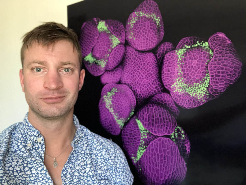
Nat Prunet
Interview by Kate Schwarting, Programs Manager
Interview by Kate Schwarting, Programs Manager
KS: What inspired you to study the cellular processes of flower formation?
NP: More than 80% of our food comes directly from plants, the vast majority of it fruits and seeds, which are parts and products of flowers, and understanding how flowers form is critical in terms of food security, and has a lot of potential applications in agronomy. Admittedly, though, one of the main reasons why I chose to study this subject is the amazing imaging possibilities developmental biology - the study of how organisms form from a single cell after fertilization – offers, whether one studies plants or animals. I fell in love with microscopy during an internship in a plant biology lab; I used a confocal microscope to take pictures and videos of pollen germinating on the pistil (the female part of the flower, which turns into a fruit after fertilization), and forming a tube that brings the male sperm cells to the ovules for fertilization. I was awed by the incredible details of life that advanced microscopy techniques let us see, and by the beauty of the images that they can generate. I have stuck to the study of flower development since, using a combination of genetics and a variety of microscopy techniques during my PhD, then focusing on a confocal microscopy approach for my postdoc, which allows me to image live flowers as they grow. Confocal microscopy is a very powerful tool to study a quintessentially dynamic process like flower development. I also really love this technique for the aesthetic qualities of the images it produces. This artistic aspect has long been an important driver for my work.
NP: More than 80% of our food comes directly from plants, the vast majority of it fruits and seeds, which are parts and products of flowers, and understanding how flowers form is critical in terms of food security, and has a lot of potential applications in agronomy. Admittedly, though, one of the main reasons why I chose to study this subject is the amazing imaging possibilities developmental biology - the study of how organisms form from a single cell after fertilization – offers, whether one studies plants or animals. I fell in love with microscopy during an internship in a plant biology lab; I used a confocal microscope to take pictures and videos of pollen germinating on the pistil (the female part of the flower, which turns into a fruit after fertilization), and forming a tube that brings the male sperm cells to the ovules for fertilization. I was awed by the incredible details of life that advanced microscopy techniques let us see, and by the beauty of the images that they can generate. I have stuck to the study of flower development since, using a combination of genetics and a variety of microscopy techniques during my PhD, then focusing on a confocal microscopy approach for my postdoc, which allows me to image live flowers as they grow. Confocal microscopy is a very powerful tool to study a quintessentially dynamic process like flower development. I also really love this technique for the aesthetic qualities of the images it produces. This artistic aspect has long been an important driver for my work.
KS: Can you describe the scientific and artistic processes you use to arrive at your images?
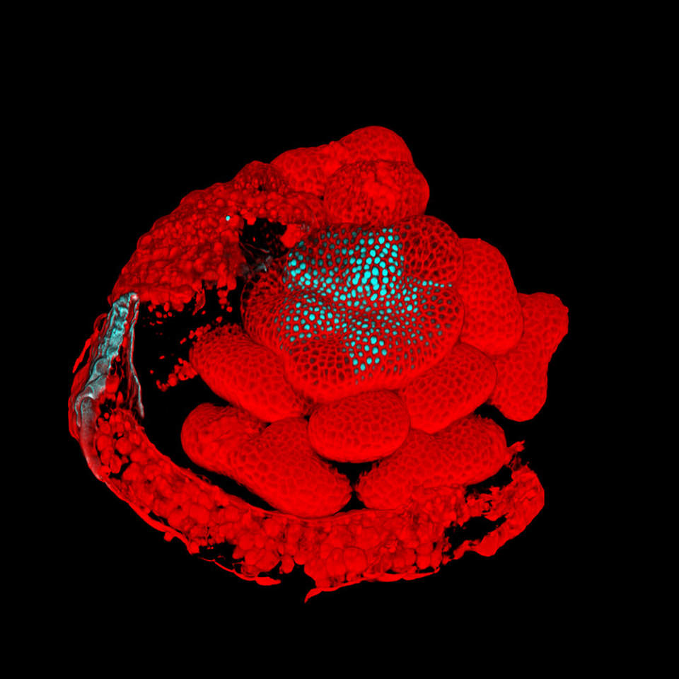
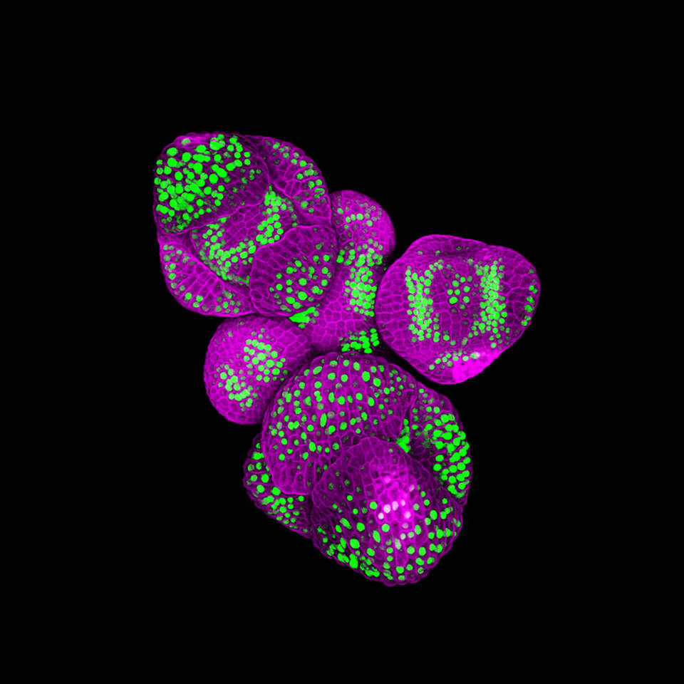
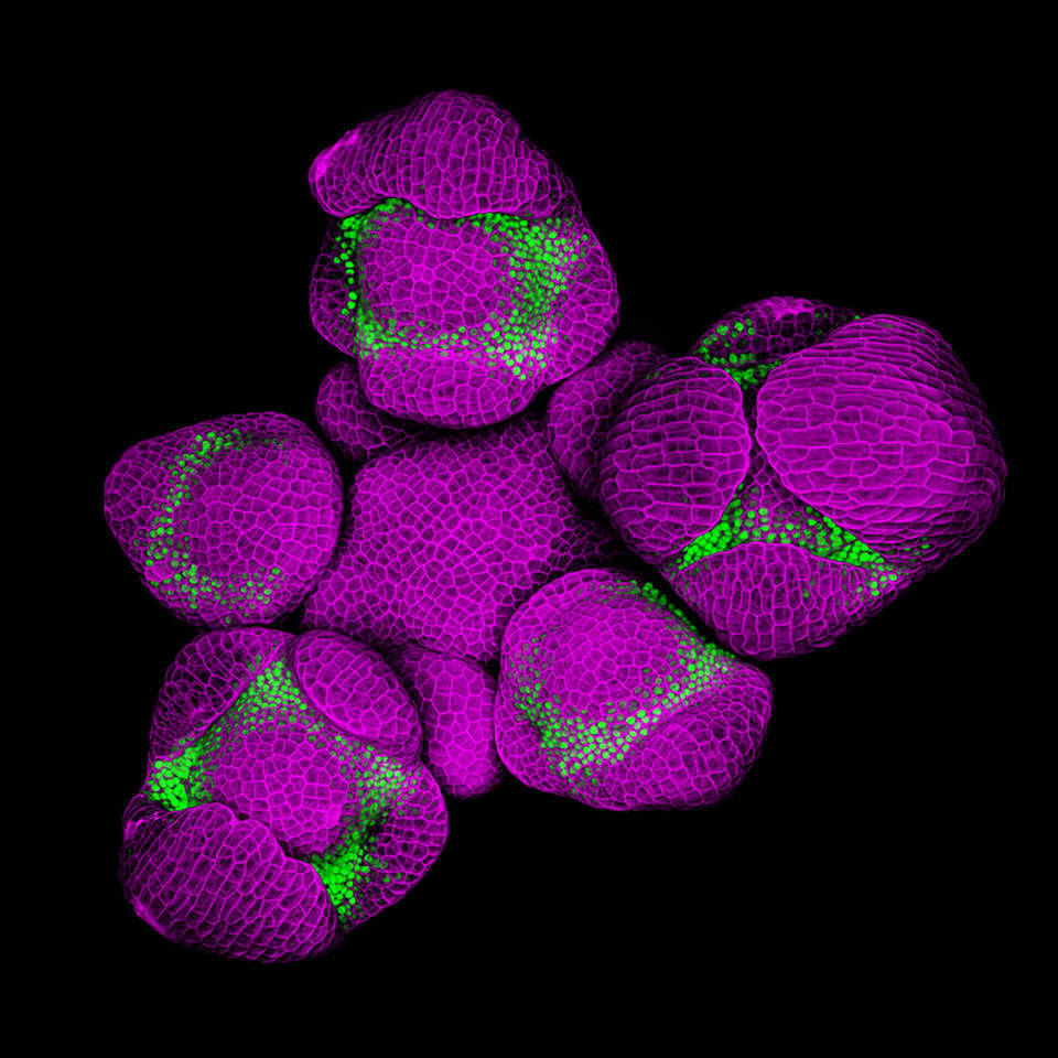
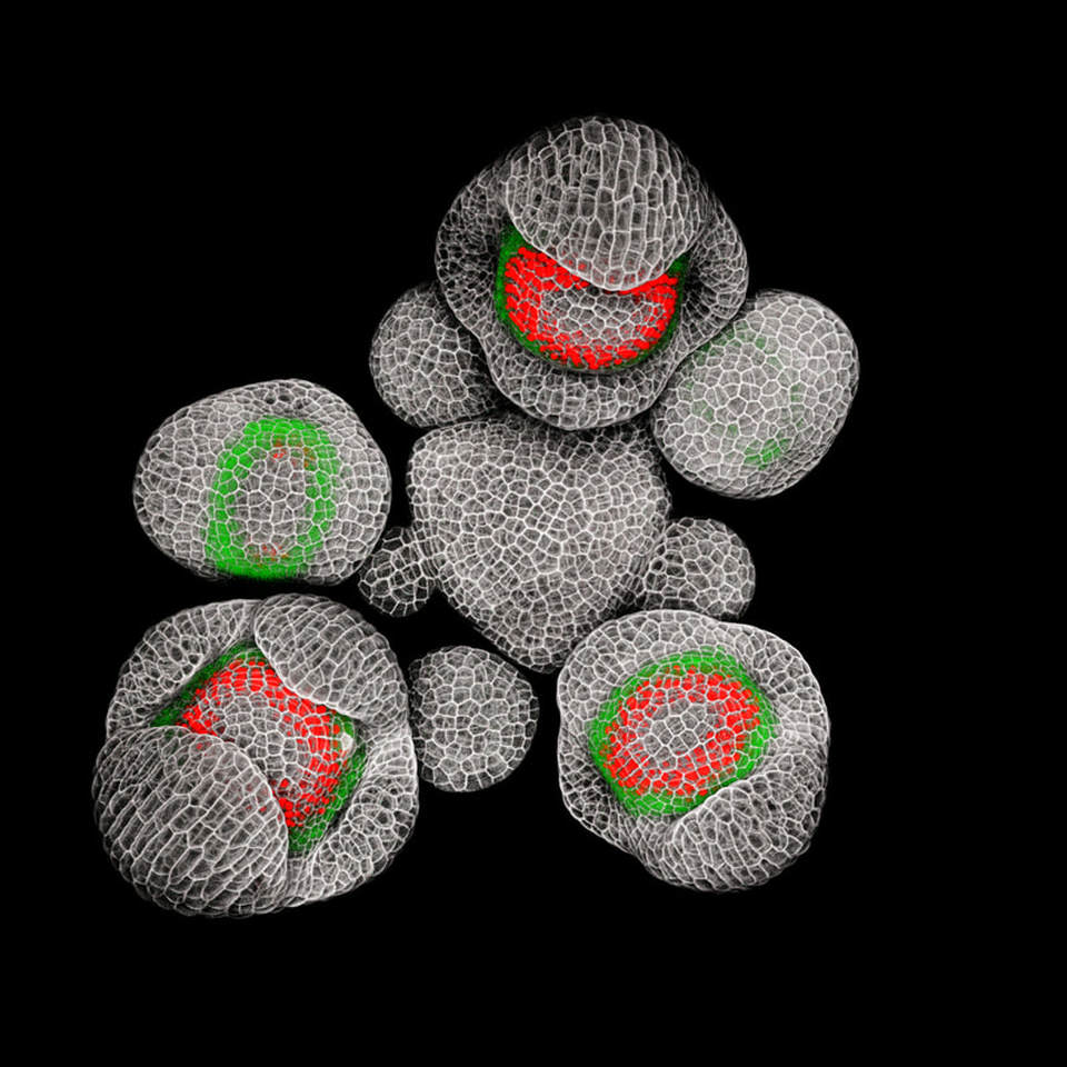
NP: My microscopy images show the tip of stems, with young flower buds forming on their flanks or flower buds on their own, with fluorescence marking the parts of the flowers where different plant genes are expressed. Every cell in an organism has the same genome. What makes cells differentiate to form a variety of tissues and organs is that they activate - or express - different subsets of their genes. To understand the cellular process of flower formation, I look at the patterns of expression of genes that influence the morphology of the flower.
The first step is to make the flowers I study fluorescent. To do that, I copy plant genes of interest and fuse them to fluorescent genes from jellyfish. I then introduce these chimeric plant/jellyfish genes back into the plant genome (this makes the plants I study genetically modified organisms, or GMOs, although they are designed for research, and not agricultural purposes). Cells that normally express specific plant genes also express the corresponding chimeric plant/jellyfish genes, which makes them fluorescent. I also use fluorescent dyes to stain the molecular “walls” that surround the cells.
To prepare these fluorescent plants for the microscope, I use thin forceps to remove fruits and flowers from the stem, and leave only the youngest flower buds that are just starting to form at the tip. This is done under a stereomicroscope, and requires really fine motor skills (caffeine is better avoided!): at the end of the dissection process, the sample is smaller than a pinhead.
I then image the tip of these stems with a confocal microscope - a type of fluorescence microscope that uses a pinhole to filter out the light that comes from out-of-focus parts of the sample; it generates optical slices of the object you study without having to physically slice it (the flowers I look at are still alive, and I can image them a few days in a row to observe their growth). Optical slices taken at different depths can then be stacked together to generate a 3D digital reconstruction of the object. Some image analysis software even let you visualize data in virtual reality!
3D digital reconstruction of flower buds
KS: What are your thoughts on the intersection of art and science?
NP: With naturalism, art and science used to go hand-in-hand, and illustration was central to the scientific process. Historically, some of the most prominent scientists, people like Ernst Haeckel or the Blaschkas, were also incredible artists. With the specialization of modern sciences came a dichotomy between science and art, but some disciplines still retain a strong artistic component. I think this is particularly the case of life sciences, for which observation drawing remains an integral part of the learning process. This artistic side is one of the reasons I have always loved, and decided to study, biology. Later, as a scientist, I have oriented my research towards microscopy because it let me keep a strong artistic component in my work. Recent technological advances have provided scientists with a wide range of techniques and instruments that produce images that are both beautiful and original- microscopy is only one of them!
I have always spent more time on the microscope, trying to get aesthetically perfect images than needed for strictly scientific purposes. Despite that, it took me a while to realize that SciArt was actually a thing. Twitter was an eye-opener for me, as I started following other scientists like Adam Summers (@Fishguy_FHL on Twitter) or Igor Siwanowicz, who take stunning images for their research, and actively promote them as art.
It is thrilling to see how dynamic the SciArt ecosystem is, and social media make it easy to connect with other people at the intersection of art and science. While I personally came to SciArt from the science side, it has been fascinating to discover the work of many talented visual, digital and multi-media artists who draw their inspiration from science!
Still Life by Andrew McKee and Shirley Watts including contributions from Nat Prunet and Steve Craig, 2016
KS: What recent projects or exhibitions have you worked on and in what types of future projects would you like to be involved?
NP: I have had some fun collaborations recently with visual artists and musicians. Some of my microscopy images were used to design the cover and artwork for the Aves Spicere album by prog-psychedelic rock band Andromaca (https://andromacaroma.bandcamp.com/releases). I also contributed to Still Life, a digital art piece by Andrew McKee and Shirley Watts, which overlaps some of my microscopy videos with time-lapses of cell division and blooming flowers by Steve Craig. Still Life was projected on a greenhouse for the Digital Nature exhibition at the Los Angeles County Arboretum in 2016.
Science has kept me too busy over the last few months to actively work on SciArt projects, but I still intend to participate to microscopy and scientific imaging competitions like the Nikon Small World or FASEB BioArt and to submit pictures to the “Infinite Potential” joint exhibition by the SciArt Ceter and the Cambridge Stem Cell Institute. I am always happy to collaborate with other artists or SciArtists!
Still Life by Andrew McKee and Shirley Watts including contributions from Nat Prunet and Steve Craig, 2016
For more of Nat's work visit his gallery or follow him on twitter @Nat_Prunet and Instagram @microscopic_farmer

